
Page 67 - IJB-9-5
P. 67
International Journal of Bioprinting 3D-printed PLA-BG composite induces angiogenesis
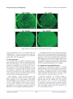
Figure 4. Adhesion of HUVECs (green) to PLA–BG disks. Scale bar: 2 mm.
adhesion and growth as well as on vascularization and As already stated before, only few studies have analyzed
wound healing [36,37] . The basic concepts of these studies are the effect of BG on vascular gene expression. It has been shown
comparable to our experimental approach and confirm the that human dental stromal cells demonstrated a higher gene
positive effect of BG on angiogenesis. expression of CD34, CD31, and KDR in 3D BG constructs .
[38]
This research group also detected osteogenic effects in the
3.5. Gene expression same scaffold . Li et al. observed a positive effect on gene
[26]
[39]
To confirm the results of the adhesion and viability assays, expression of FGF (fibroblast growth factor), VEGF and KDR
gene expression analyses of the two endothelial markers in HUVECs after being seeded on BG–PVA scaffolds.
CD31 and KDR (kinase insert domain receptor) were
performed. Considering the results from this study as 3.6. Angiogenesis assay (Matrigel assay)
well as from the studies regarding osteogenesis, one can To analyze the differentiation capacity of PLA-BG
conclude that the lowest concentration of BG has no effect scaffolds, Matrigel assays were performed to quantify tube
on osteogenic or endothelial cells. The group with the formation [33,40] . This assay is easy to perform, easy to analyze
lowest BG concentration of 5% was excluded from further and quantify, and highly reproducible. Another advantage
studies. Only PLA with BG concentrations from 10% and is that endothelial cells attach within 1 h and start to form
20% were used for the following experiments. tubes as well as cell–cell contacts within 12–24 h .
[41]
Gene expression of both endothelial markers was Exemplary illustrations of HUVECs seeded in
enhanced in both PLA–10%BG and PLA–20%BG groups Matrigel® and incubated with conditioned medium
when compared to pure PLA-scaffolds. PLA–20%BG from the three PLA–BG scaffolds (0%, 10%, and 20%
showed the highest expression of these endothelial markers BG) are shown in Figure 7A. Figure 7B demonstrates
(Figure 6). KDR expression seems to be lower on day 4 the quantitative analysis showing that all three analyzed
on PLA–20%BG compared to PLA–10%BG; however, components (number of junctions, number of segments,
the difference is not statistically significant, whereas the and total length) increased with BG content in the
difference to PLA pure group is statistically significant. 3D-printed scaffolds (Figure 7).
Volume 9 Issue 5 (2023) 59 https://doi.org/10.18063/ijb.751