
Page 69 - IJB-9-5
P. 69
International Journal of Bioprinting 3D-printed PLA-BG composite induces angiogenesis
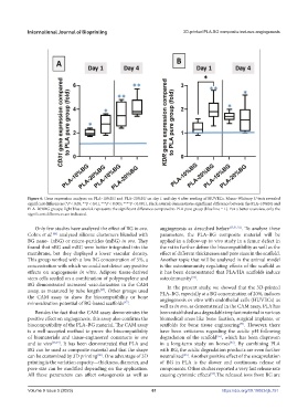
Figure 6. Gene expression analyses on PLA–10%BG and PLA–20%BG on day 1 and day 4 after seeding of HUVECs. Mann–Whitney U tests revealed
significant differences (*P < 0.05, **P < 0.01, ***P < 0.005; ****P < 0.001). Black asterisk demonstrates significant difference between the PLA–10%BG and
PLA–20%BG groups; light blue asterisk represents the significant difference compared to PLA pure group (blue line = 1). For a better overview, only the
significant differences are indicated.
Only few studies have analyzed the effect of BG in ovo. angiogenesis as described before [25,51,52] . To analyze these
[46]
Cohrs et al. analyzed silicone elastomers blended with parameters, the PLA–BG composite material will be
BG nano- (nBG) or micro-particles (mBG) in ovo. They applied in a follow-up in vivo study in a femur defect in
found that nBG and mBG were better integrated into the the rat to further define the biocompatibility as well as the
membrane, but they displayed a lower vascular density. effect of different thicknesses and pore sizes in the scaffold.
This group worked with a low BG concentration of 5%, a Another topic that will be analyzed in the animal model
concentration with which we could not detect any positive is the osteoimmunity regulating effects of the scaffold as
effects on angiogenesis in vitro. Adipose tissue-derived it has been demonstrated that PLA/HA scaffolds induce
[53]
stem cells seeded on a combination of polypropylene and osteoimmunity .
BG demonstrated increased vascularization in the CAM In the present study, we showed that the 3D-printed
[27]
assay, as measured by tube length . Other groups used PLA–BG, especially at a BG concentration of 20%, induces
the CAM assay to show the biocompatibility or bone angiogenesis in vitro with endothelial cells (HUVECs) as
[47]
mineralization potential of BG-based scaffolds .
well as in ovo, as demonstrated in the CAM assay. PLA has
Besides the fact that the CAM assay demonstrates the been established as a degradable implant material in various
positive effect on angiogenesis, this assay also confirms the biomedical areas like bone fixation, surgical implants, or
biocompatibility of the PLA–BG material. The CAM assay scaffolds for bone tissue engineering . However, there
[54]
is a well-accepted method to prove the biocompatibility have been criticisms regarding the acidic pH following
of biomaterials and tissue-engineered constructs in ovo degradation of the scaffold , which has been disproven
[55]
and in vivo [48,49] . It has been demonstrated that PLA and in a long-term study on horses . By combining PLA
[56]
BG can be used as composite material and that the shape with BG, the acidic degradation products are even further
can be customized by 3D printing . One advantage of 3D neutralized . Another positive effect of the encapsulation
[50]
[55]
printing is the variation capacity—thickness, diameter, and of BG in PLA is the slower and continuous release of
pore size can be modified depending on the application. components. Other studies reported a very fast release rate
All these parameters can affect osteogenesis as well as causing cytotoxic effects .The released ions from BG are
[57]
Volume 9 Issue 5 (2023) 61 https://doi.org/10.18063/ijb.751