
Page 162 - IJB-9-6
P. 162
International Journal of Bioprinting 3D printing in gastroenterology
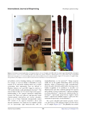
Figure 2. Illustrations of several applications of 3D-printed objects. (A) An abdominal cavity filled with 3D-printed organs that match the radiological
[35]
[60]
density in real CT scan ; (B) 3D-printed steerable instruments that improve flexibility in laparoscopy ; (C) A 3D-printed model of esophageal
submucosal tumor (green) to be resected using endoscopic submucosal dissection and its adjacent anatomy. Figure 2A–B are reprints of original images
with permission (The images are licensed under Creative Commons Attribution 4.0 International License).
[28]
and patients, such as teaching, training, and counseling. lymphadenectomy in five patients . Young surgeons
For example, an anorectal fistula is intractable to assess could refer to the spatial relationships when reviewing
because of its anatomical winding routes and complex operative videos of laparoscopic right hemicolectomy,
connections. Most importantly, they are difficult to which the author believed could shorten learning curves.
illustrate. Sahnan et al. used MRI images to construct a Similar recognition of its usefulness in education and
3DP model of fistula canals and adjacent structures . The simulation has also been achieved among both experts
[27]
[29]
authors believed that real 3D models improved surgeons’ and residents in bowel anastomosis , tracheoesophageal
[30]
understanding of the complex anatomical relationship prosthesis placement , laparoscopic pyloromyotomy for
[31]
between the fistula and sphincter and provided better neonates , laparoscopic preperitoneal inguinal hernia
[33]
[32]
clinician–patient relationships and medical education. repair , laparoscopic bariatric surgery , and cystic duct
[34]
Hojo et al. of Tokyo University retrospectively created variations in laparoscopic cholecystectomy .
3DP models of the superior mesenteric artery and For a purer educative and research purpose, Anwari
superior mesenteric vein based on the surgeries carried et al. developed a 3DP anthropomorphic phantom based
out for laparoscopic right hemicolectomy with D3 on CT images (Figure 2A) . The phantom was created
[35]
Volume 9 Issue 6 (2023) 154 https://doi.org/10.36922/ijb.0149