
Page 167 - IJB-9-6
P. 167
International Journal of Bioprinting 3D printing in gastroenterology
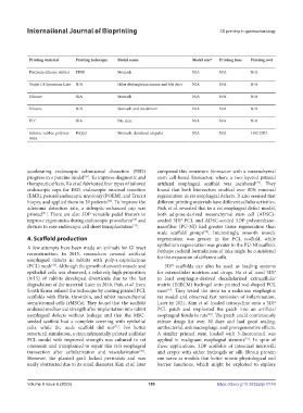
Table 2. 3D printing for endoscopic operation, education, and simulation
Author Year Application Image Data Image processing Output 3D printing software Printing machine Printing material Printing technique Model name Model size* Printing time Printing cost
source format software format
Lee et al. [65] 2018 Endoscopic biopsy CT DICOM 3D Slicer v.4.5.0 STL Netfabb professional Clone S270 & Clone Platinum silicone rubber FDM Stomach N/A N/A N/A
v.5 K300
Yang et al. [61] 2018 ERCP CT/MRI DICOM Mimics Innovation STL N/A ProJet 4500 Visijet C4 Spectrum Core N/A Hilar cholangiocarcinoma and bile duct N/A N/A N/A
Suite v17.0
Lee et al. [64] 2019 Endoscopic CT DICOM 3D Slicer v.4.5.0 STL Netfabb professional Form 2 Silicone SLA Stomach N/A N/A N/A
hemostasis v.5
Kwon et al. [67] 2020 ERCP CT N/A In-house software STL MeshLab and 3DM Tough-3.6 Silicone N/A Stomach and duodenum N/A N/A N/A
MeshMixer
Dhir et al. [109] 2015 EUS-guided MRI N/A N/A N/A N/A Viper SI2 PLC SLA Bile duct N/A N/A N/A
biliary drainage
Holt et al. [68] 2018 Endoscopic N/A N/A N/A N/A Solid Works 2014 Connex 260v Silicone rubber, polymer Polyjet Stomach, duodenal ampulla N/A N/A 1482 USD
ampullectomy resin
Abbreviations: CT, computed tomography; DICOM, digital imaging and communications in medicine; ERCP, endoscopic retrograde cholangiopan-
creatography; FDM, fused deposition modeling; MRI, magnetic resonance imaging; PLC, polycarbonate; SLA, stereo lithography appearance; STL,
stereolithography; *refers to the percentage of lifesize organs.
accelerating endoscopic submucosal dissection (ESD) compared this omentum bioreactor with a mesenchymal
progress in a porcine model . To improve diagnostic and stem cell-based bioreactor, where a two-layered printed
[69]
therapeutic effects, Ko et al. fabricated four types of tailored artificial esophageal scaffold was incubated . They
[77]
endoscopic caps for ESD, endoscopic mucosal resection found that both bioreactors enabled over 80% mucosal
(EMR), peroral endoscopic myotomy (POEM), and Trucut regeneration in rat esophageal defects. It also seemed that
biopsy, and applied them in 39 patients . To improve the different printing materials have different cellular activities.
[70]
adenoma detection rate, a sideoptic-enhanced cap was Park et al. revealed that in a rat esophageal defect model,
printed . There are also 3DP versatile pedal fixators to both adipose-derived mesenchymal stem cell (ADSC)-
[71]
improve ergonomics during endoscopic procedures and seeded 3DP PCL and ADSC-seeded 3DP polyurethane-
[72]
devices to ease endoscopic cell sheet transplantation . nanofiber (PU-Nf) had greater tissue regeneration than
[73]
nude scaffold groups . Interestingly, smooth muscle
[78]
4. Scaffold production regeneration was greater in the PCL scaffold, while
epithelium regeneration was greater in the PU-Nf scaffold.
A few attempts have been made on animals for GI tract Perhaps cocktail formulations of inks might be considered
reconstruction. In 2015, researchers covered artificial for the expansion of different cells.
esophageal defects in rabbits with poly-ε-caprolactone
[74]
(PCL) mesh . Although the growth of smooth muscle and 3DP scaffolds can also be used as loading systems
epithelial cells was observed, a relatively high proportion for extracellular matrices and drugs. Ha et al. used 3DP
(9/15) of rabbits developed diverticula due to the fast to load esophagus-derived decellularized extracellular
degradation of the material. Later in 2016, Park et al. from matrix (EdECM) hydrogel onto printed rod-shaped PCL
South Korea refined the technique by coating printed PCL stent . They tested the stent in a radiation esophagitis
[79]
scaffolds with fibrin, thrombin, and rabbit mesenchymal rat model and observed fast remission of inflammation.
stem/stromal cells (rMSCs). They found that the scaffold Later in 2021, Kim et al. loaded tetracycline onto a 3DP
endured mechanical strength after implantation into rabbit PCL patch and implanted the patch into an artificial
esophageal defects without leakage and that the MSC- esophageal fistula in rats . The patch could continuously
[80]
seeded scaffold had a complete covering with epithelial release drugs for over 30 days and had good sealing,
cells, while the nude scaffold did not . For better antibacterial, antimacrophage, and proregenerative effects.
[75]
structural simulation, a circumferentially printed acellular A similar printed stent loaded with 5-fluorouracil was
PCL model with improved strength was cultured in rat applied in malignant esophageal stenosis . In spite of
[81]
omentum and transplanted to repair this rat’s esophageal these applications, 3DP scaffolds of intestinal microvilli
transection after cellularization and vascularization . and crypts with either hydrogels or silk fibroin protein
[76]
However, the planted graft lacked peristalsis and was can serve as models that better mimic physiological and
easily obstructed due to its small diameter. Kim et al. later barrier functions, which might be exploited to explore
Volume 9 Issue 6 (2023) 159 https://doi.org/10.36922/ijb.0149