
Page 338 - IJB-9-6
P. 338
International Journal of Bioprinting 3D printing and bioprinting in urology
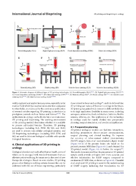
Figure 5. Schematic diagram of different types of 3D printing technologies. (A) Stereolithography (SLA) [83,84] . (B) Digital light processing (DLP) [85,86] .
(C) Fused deposition modeling (FDM) . (D) Direct ink writing (DIW) [28-30] . (E) Material jetting (MJ) . (F) Binder jetting (BJ) [34,35] . (G) Selective laser
[8]
[10]
sintering (SLS) [37,87] . (H) Selective laser melting (SLM) [38,88,89] .
widely explored and applied in many areas, especially in the those related to bone and cartilage , and it is believed that
[9]
medical field which has received more attention compared 3D printing can make a difference in urology in the future.
to other fields, as evidenced by the continuous publication 3D printing has gained wide interest in different fields due
of research articles regarding 3D printing in medicine in to its high application flexibility in many areas, including
prestigious journals such as Nature and Science [42-47] . The aerospace, automotive, food, architecture, batteries, flexible
applications in urology can be divided into two main areas: sensors, robotics, etc. The application of this technology
3D printing and bioprinting. The working environment in urology could be mainly divided into preoperative
of the printing method determines whether it is available planning, surgical simulation, and preclinical application.
to process cell-loaded bioinks. Therefore, 3D printing
technologies (including SLA, FDM, BJ, SLS, and SLM) 4.1. Preoperative planning
are used to process non-cellular urological samples, and 3D-printed urological models can facilitate interactions,
3D bioprinting technologies (including DLP, DIW, and including preoperative doctor–patient communication,
MJ) are used to fabricate biological scaffolds with specific surgical planning, and clinical teaching. To improve
biological functions. the accuracy of robot-assisted radical prostatectomy
(RARP) surgery, Saba et al. reconstructed a digital model
4. Clinical applications of 3D printing in (Figure 6A-ii) of the prostate lesion site based on the
urology patient’s prostate MRI data (Figure 6A-i) and obtained the
3D-printed model (Figure 6A-iii) before the surgery .
[48]
Urological disorders seriously affect human health, some of The results demonstrated that the preoperative 3D-printed
which can be managed with standard treatments that can sample could provide urologists with more accurate and
alleviate patient suffering, but many are in dire need of new effective information for RARP surgery compared to
therapeutic strategies. Based on some studies, 3D printing continuous 2D MRI data, as shown in Figure 6A. Chandak
gives a silver lining in tackling certain diseases that can be et al. employed 3D printing to assist in the understanding
addressed with the use of tissue engineering, particularly of RARP surgery, indicating that visualization via 3D
Volume 9 Issue 6 (2023) 330 https://doi.org/10.36922/ijb.0969