
Page 340 - IJB-9-6
P. 340
International Journal of Bioprinting 3D printing and bioprinting in urology
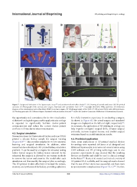
Figure 7. Design and fabrication of the laparoscopic trocar and extraluminal anti-reflux diodes . (A) Drawing of cannula and trocar. (B) 3D-printed
[90]
[55]
cannulas. (C) Photograph of the cannula after surgery. Reprinted with permission from ref. . Copyright 2022 John Wiley and Sons. (D) Schematic
[90]
diagram of the extraluminal anti-reflux diode (EAD) in urinary organs. (E) Working principle of the EAD. (F) 3D-printed EADs with different shapes.
(E) Working principle. (F) Different shapes. Reprinted with permission from ref. under the Creative Commons Attribution 4.0 International License.
[55]
the opportunity and convenience for in vitro visualization for a fully immersive experience in conducting a surgery.
of diseased urological organs, and its application in urology As shown in Figure 6E, the actual surgery and simulated
is expected to significantly facilitate doctor–patient images are displayed on the left and right, respectively .
[52]
communication and reduce the current doctor–patient In summary, the application of 3D printing in urology can
conflicts in China due to miscommunication. help improve urologists’ surgical skills, enhance surgical
protocols, increase surgical success, and predict surgical
4.2. Surgical simulation outcomes based on model simulation results.
Researchers from the University of California Irvine (USA)
printed a silicone kidney sample for surgical training 4.3. Preclinical application
(Figure 6D) . This provides samples for in vitro anatomy Here, some applications of 3D-printed medical devices
[51]
learning and surgical simulation. In addition, other for urology were reported. del Junco et al. designed and
researchers have developed a 3D-printed kidney simulation fabricated laparoscopic punctures and ureteral stents using
platform. It can be used by urologists for rehearsal testing CAD software and 3D printing technology, and in vivo
before formal surgery for kidney tumor removal. In the puncture experiments in pigs showed that the resulting
rehearsal, the surgeon is allowed to use various methods devices are feasible and could be used in clinical settings
to remove the tumor and examine the model after each in the future . Buote et al. created and trained a variety of
[53]
simulation and thus modify the surgical plan accordingly. 3D-printed PLA scaffolds, and the surgical results showed
The 3D-printed models effectively simulated the texture, that the use of their stents was successful, and there were
anatomy, and perfusion of the kidney, providing a platform no postoperative complications (Figure 7A–C) . Further,
[90]
Volume 9 Issue 6 (2023) 332 https://doi.org/10.36922/ijb.0969