
Page 345 - IJB-9-6
P. 345
International Journal of Bioprinting 3D printing and bioprinting in urology
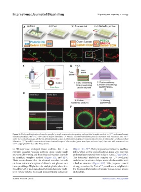
Figure 10. Design and fabrication of vascular samples by single-nozzle extrusion printing and sacrificial template method (A, B) , and coaxial-nozzle
[71]
extrusion printing (C–F) . (A) Flow chart of sample fabrication. (B) Vascular samples with different patterns. Reprinted with permission from ref.
[71]
[74]
under the Creative Commons Attribution 4.0 International License. (C) Schematic diagram of coaxial-nozzle structure. (D) Flow chart of tubular scaffold
fabrication. (E) Top and (F) cross-sectional views of stained images of tube samples (green, inner layer; red, outer layer). Reprinted with permission from
ref. . Copyright 1999–2023 John Wiley & Sons.
[74]
in 3D-bioprinted urological tissue scaffolds. Lin et al. (Figure 10C–F) . Their proposed coaxial nozzle has three
[74]
prepared complex vascular patterns using single-nozzle inlets, which are the central material, inner layer material,
extrusion 3D printing and then obtained vascular channels and outer layer material from inside to outside (Figure 10C).
by sacrificial template method (Figure 10A and B) . The fabricated triple-layer samples are UV-crosslinked
[71]
Their results showed that the obtained vascular channels and cleaned to obtain a bilayer material tube scaffold with
exhibited active reabsorption of albumin and glucose over a hollow structure (Figure 10E). This proposed coaxial
time, providing a 3D platform for studying kidney function. strategy conceived by Pi et al. provides new insights into
[74]
In addition, Pi et al. designed and fabricated annular multi- the design and fabrication of tubular tissues such as ureters
layer tubular samples by coaxial-nozzle printing technology and urethra.
Volume 9 Issue 6 (2023) 337 https://doi.org/10.36922/ijb.0969