
Page 343 - IJB-9-6
P. 343
International Journal of Bioprinting 3D printing and bioprinting in urology
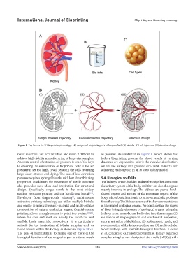
Figure 8. Key factors for 3D bioprinting in urology: (A) design and bioprinting of a kidney scaffold, (B) bioinks, (C) cell types, and (D) structure design.
result in serious ink accumulation and make it difficult to as possible. As illustrated in Figure 8, which shows the
achieve high-fidelity manufacturing of large-size samples. kidney bioprinting process, the blood vessels of varying
Accurate control of extrusion air pressure is one of the keys diameter are expected to mimic the vascular distribution
to ensuring the survival rate of bioprinted cells; if the air within the kidney and provide structural mimicry for
pressure is set too high, it will result in the cells receiving achieving reabsorption in an in vitro kidney model.
large shear stresses and dying. The use of low extrusion
pressure requires hydrogel bioinks with low shear thinning 5.4. Urological scaffolds
properties. In addition, the innovation of nozzle structure The kidneys, ureter, bladder, and urethra together constitute
also provides new ideas and inspiration for structural the urinary system of the body, and they are also the organs
design. Specifically, single nozzle is the most widely mainly involved in urology. The kidneys are paired lentil-
used in extrusion printing and can handle one bioink . shaped organs and are one of the important organs of the
[81]
Developed from single-nozzle printing , multi-nozzle body, whose basic function is to remove metabolic products
[3]
extrusion printing technology can utilize multiple bioinks from the body. The kidneys are one of the key representatives
and enable to mimic the multi-material and multi-cellular of bioprinted urological organs. We conclude that the stages
composition of natural urological organs. Coaxial-nozzle of bioprinting development of urological organs, using the
printing allows a single nozzle to print two bioinks [75,82] , kidneys as an example, can be divided into three stages: (1)
where the core and shell are usually the sacrificial and realization of simple physical and mechanical properties,
scaffold body materials, respectively. It is particularly such as urination of the kidney; (2) replacement, repair, and
suitable for the fabrication of tubular samples, such as reconstruction of the kidney urethra; and (3) multi-cellular
blood vessels within the kidney, as shown in Figure 8C-ii. bionic kidneys with multiple biological functions. Lawlor
The goal of bioprinting is to mimic one or more of the et al. conducted extrusion bioprinting of kidney organoid
biological functions of a urological organ in vitro as much samples using human pluripotent stem cells (hPSCs) with
Volume 9 Issue 6 (2023) 335 https://doi.org/10.36922/ijb.0969