
Page 551 - IJB-9-6
P. 551
International Journal of Bioprinting 3D printing of PCL-ceramic composite scaffolds
A
B C
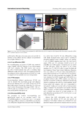
Figure 2. (A) Direct-write 3D printing custom equipment for scaffold fabrication. (B) 3D computer-aided design model of the ceramic composite scaffold.
(C) 3D-printed composite ceramic scaffold.
performed to eliminate extraneous pores and provide the in a sterile fume hood for 30 min, followed by rinsing
porosity of the scaffold. Porosity analysis was performed with sterile deionized water (twice) and 1× Dulbecco’s
on a sample of three (n = 3). phosphate-buffered saline (DPBS). Before cell seeding,
1 mL of DMEM supplemented with 10% fetal bovine
2.4.4. X-ray diffraction (XRD)
serum (FBS) and 1% antibiotics (10,000 units/mL of
The crystallography and phases of CMP were examined penicillin and 10,000 μg/mL of streptomycin) was added
using a Bruker AXS D8 Discover X-ray diffractometer to each well plate, which was placed in cell culture
with Cu-K radiation. The XRD studies were carried incubator for 3 h. NIH/3T3 cells, a mouse fibroblast cell
out with a locked-coupled scan with a scanning range line (American Tissue Type Culture Collection, Manassas,
(diffraction angle, 2θ) of 15°–60°. The instrument was run VA), were cultured in a 75 cm culture flask and kept in a
2
in continuous mode, with increments of 0.0146° for 2 min. tissue culture incubator at 37°C and a 5% CO atmosphere.
2
An experiment was carried out at room temperature. Every 2 days, the culture medium was changed. The cells
2.4.5. FT-IR analysis were separated by 0.025% trypsin and 0.01% EDTA in a
phosphate-buffered saline (PBS) solution once they had
Fourier-transform infrared spectroscopy (FT-IR) was reached around 90% confluence, followed by transferring
used to identify the functional groups and chemical them to a centrifuge tube with the culture medium. Before
interactions between PCL and CMP. Varian 670 FT-IR being seeded into samples, cells were resuspended in
Spectrophotometer (Varian, Inc., Palo Alto, CA, USA) was new growth media and counted with a hemocytometer
used to detect the spectra in the range of 4000 – 400 cm using a Countess II Automated Cell Counter (Thermo
−1
TM
with 64 scans at a resolution of 4 cm . A total of 5 scans Fisher Scientific). A 50 μL aliquot of medium containing
−1
were performed for each spectrum.
cells (∼50,000) was placed on printed samples (n = 3)
2.4.6. Biocompatibility study and cultured in an incubator (37°C, 5% CO ) for 1, 2, and
2
3 days, respectively.
The procedure to conduct biocompatibility was
implemented based on our previous work . The Alamar blue (AB) colorimetric assay was used
[29]
3D scaffold samples (n = 3) were cut (1 cm × 1 cm), glued to measure the viability of 3T3 cells after growth on
with Surgical Silicone Adhesive, Kwik-SilTM, and attached substances. Cell culture medium was collected from each
to 24-well plates. Samples were sterilized with 95% ethanol incubated sample and stored for toxicity study at a specific
Volume 9 Issue 6 (2023) 543 https://doi.org/10.36922/ijb.0196