
Page 554 - IJB-9-6
P. 554
International Journal of Bioprinting 3D printing of PCL-ceramic composite scaffolds
3.4. Porosity of scaffolds for facilitating the diffusion of nutrition, allowing cell
The pore size and porosity of the 3D-printed scaffolds migration, accelerating cell proliferation, and enabling
[66-68]
were calculated and are presented in Table 2. The virgin vascularization . Thus, our high-porosity scaffolds
polymer PMC-0 had the largest pore size (~245 μm) and provide diffusion and release pathways of biological
the highest porosity (50%), respectively. However, as the molecules and nutrients for cellular migration and
ceramic content within the scaffold increased, there was proliferation [69,70] .
a reduction in both the pore size and porosity. This can 3.5. Hydrophilicity behavior of scaffolds
be attributed to the increase in ceramic loading within
the polymer composite that leads to higher viscosities The surface wettability of the scaffolds, which affects cell
of the 3D-printed slurries. This finding correlates well proliferation and protein absorption, can be determined by
with the rheological behavior of the PMC suspension the water contact angle. Hydrophilicity plays a crucial role
as shown in Figure 3, wherein higher microparticle in cell interaction within the scaffold. The hydrophilicity
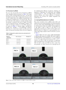
loading has revealed a non-Newtonian behavior. of the PMC-0, PMC-5, PMC-10, and PMC-15 was
However, it is noteworthy to point out that pore sizes analyzed by measuring the incident contact angle at two
above 150 μm and porosities above 40% are conducive different durations (initial at 3 s and equilibrium 90 s)
using a drop shape analyzer (KRUSS-DSA25E) as shown
Table 2. Comparative analysis of pore size and porosity for in Figure 7.
PMC scaffolds Figure 8 shows the water contact angle measurements
of polymer and composite scaffolds. The contact angle for
Material Pore size (μm) Porosity (%)
composition our blend of PMC-0 was around 94.31 ± 3.21° as compared
[71]
PMC-0 245.5±20.5 50.61±0.34 to pure PCL reported in the literature at 109.2 ± 4.1° .
PMC-5 234.3±22.4 48.32±0.23 However, the PMC-5, PMC-10, and PMC-15 composites
had consistently lower contact angles (PMC-5: 74.5 ± 2.23°;
PMC-10 222.8±23.2 45.54±0.71 PMC-10: 68.9 ± 2.15°; and PMC-15: 67.8 ± 2.03°). The
PMC-15 213.4±18.7 42.34±0.56 incorporation of CMPs increased the hydrophilicity of
Figure 7. Water contact angle measurement of different contents of PMCs scaffolds.
Volume 9 Issue 6 (2023) 546 https://doi.org/10.36922/ijb.0196