
Page 240 - v11i4
P. 240
International Journal of Bioprinting Fine collagen scaffold for osteogenesis
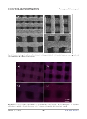
Figure 8. Microscale SEM images of crosslinked scaffolds. (A) Sample 1; (B) Sample 2; (C) Sample 3; (D) Sample 4. The scale bar shown is applicable to all
panels. Abbreviation: SEM: Scanning electron microscopy.
Figure 9. Microscale images of scaffolds seeded with MSCs and stained with DAPI and actin. (A) Sample 1; (B) Sample 2; (C) Sample 3; (D) Sample 4. The
scale bar shown is applicable to all panels. Abbreviations: DAPI: 4’6-diamidino-2-phenylindole; MSCs: Mesenchymal stem cells.
Volume 11 Issue 4 (2025) 232 doi: 10.36922/IJB025140116