
Page 310 - v11i4
P. 310
International Journal of Bioprinting 3D scaffold prevents tendon ossification
and SF–HPC bioinks gradually decreased with increasing HPC into SF enhanced cross-linking and promoted a more
shear rate, indicating favorable shear-thinning behavior. stable network within the bioink. In conclusion, excellent
Shear-thinning capability is one of the core requirements rheological properties are crucial for successful extrusion-
for extrusion-based 3D bioprinting. At the high shear rate based 3D bioprinting, as they directly affect the bioinks’
41
near the printer nozzle, viscosity decreased significantly, extrusion controllability, structural fidelity, cell viability,
enabling the bioinks to pass through the micro-nozzle and ultimately the functionality of engineered tissues. 43
with low resistance, thereby preventing nozzle clogging
due to high viscosity. Upon exiting the nozzle, the shear 3.2. Three-dimensional bioprinting of tissue-
rate abruptly dropped, and viscosity rapidly recovered, engineered Achilles tendon scaffolds and
enhancing scaffold shape stability and preventing structural bending tests
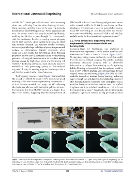
collapse or deformation. Equally important, when Extrusion-based 3D bioprinting was employed to
using cell-laden bioinks for 3D printing, shear-thinning fabricate tissue-engineered Achilles tendon scaffolds with
properties enable smooth extrusion under lower printing dimensions of 2 mm × 3 mm × 15 mm (Figure 2A–C).
pressure, reducing cell membranes or extracellular matrix During the printing process, the bioink extruded smoothly
damage caused by high shear stress and improving cell from the nozzle without clogging. The printed scaffolds
viability. Following extrusion, rapid viscosity recovery maintained structural integrity with no observable
42
immobilizes cells, preventing gravity or flow-induced deformation or collapse, demonstrating excellent printing
migration during the molding stage and ensuring uniform fidelity. Bending tests revealed that the scaffolds could be
bent to 180° without fracture and fully recovered to their
cell distribution within the printed structure.
original shape after unloading (Figure 2D1–D3). SF–HPC
The frequency–modulus curve (Figure 1B) showed that scaffolds showed no fracture during bending, indicating
the G’ and G’’ of both SF and SF–HPC bioinks remained superior toughness and ductility. Complete shape recovery
relatively stable with increasing frequency. Notably, the G’ after bending further confirmed their appropriate elastic
of both bioinks exceeded their respective G’’, indicating modulus. Previous studies have demonstrated that scaffold
that both formulations exhibited stable gel-like behavior. toughness, ductility, and elastic modulus are critical factors
Furthermore, the G’ of SF–HPC bioinks was higher than for tendon injury repair. Specifically, the Achilles tendon
44
that of SF bioinks, suggesting that the incorporation of undergoes significant tension during physical activities
Figure 2. Fabrication and bending test of 3D bioprinted tissue-engineered Achilles tendon scaffolds. (A) Digital design model prior to bioprinting. (B) 3D
bioprinting process. (C) Macroscopic view of the printed scaffold. (D1–D3) Sequential bending test demonstrating elastic recovery (180° bending angle)
and preserved structural integrity (no fractures observed). Abbreviation: 3D, three-dimensional.
Volume 11 Issue 4 (2025) 302 doi: 10.36922/IJB025210203