
Page 325 - v11i4
P. 325
International Journal of Bioprinting Nozzle geometry for enhanced cell viability
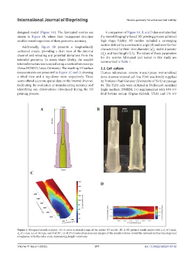
designed model (Figure 1A). The fabricated nozzles are A comparison of Figure 1A, B, and D demonstrates that
shown in Figure 1B, where their transparent structure the stereolithography-based 3D printing process achieved
enables visual inspection of their geometric accuracy. high shape fidelity. All nozzles included a converging
section defined by a contraction angle (θ) and were further
Additionally, Figure 1B presents a longitudinally
sectioned nozzle, providing a clear view of the internal characterized by their inlet diameter (d ), outlet diameter
1
(d ), and land length (LL). The values of these parameters
channel and revealing any potential deviations from the for the nozzles fabricated and tested in this study are
2
intended geometry. To assess shape fidelity, the nozzle’s summarized in Table 1.
internal structure was scanned using a confocal microscope
(Leica DCM3D, Leica, Germany). The resulting 3D surface 2.2. Cell culture
measurements are presented in Figure 1C and D, showing Human telomerase reverse transcriptase immortalized
a tilted view and a top–down view, respectively. These bone marrow stromal cell line Y201 was kindly supplied
scans offered accurate spatial data on the internal channel, by Professor Paul Genever (University of York) at passage
facilitating the evaluation of manufacturing accuracy and 84. The Y201 cells were cultured in Dulbecco’s modified
identifying any deformations introduced during the 3D Eagle medium (DMEM, 1×) supplemented with 10% v/v
printing process. fetal bovine serum (Sigma-Aldrich, USA) and 1% v/v
Figure 1. Designed nozzle analysis. (A) A cross-sectional image of the nozzle 3D model. (B) A 3D-printed nozzle model with a d of 3 mm,
1
d of 1 mm, LL of 10 mm, and θ of 20°. (C & D) Confocal microscopy images of the nozzle interior reveal the internal surface topology and
2
roughness, with the color scale representing height variations.
Volume 11 Issue 4 (2025) 317 doi: 10.36922/IJB025190182