
Page 327 - v11i4
P. 327
International Journal of Bioprinting Nozzle geometry for enhanced cell viability
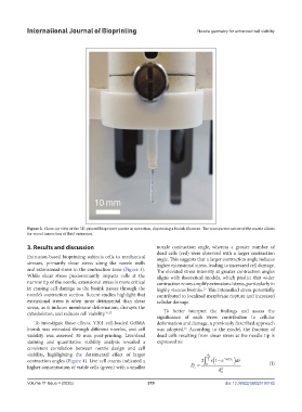
Figure 2. Close-up view of the 3D-printed bioprinter nozzle in operation, dispensing a bioink filament. The transparent nature of the nozzle allows
for visual inspection of fluid extrusion.
3. Results and discussion nozzle contraction angle, whereas a greater number of
dead cells (red) were observed with a larger contraction
Extrusion-based bioprinting subjects cells to mechanical angle. This suggests that a larger contraction angle induces
stresses, primarily shear stress along the nozzle walls higher extensional stress, leading to increased cell damage.
and extensional stress in the contraction zone (Figure 3). The elevated stress intensity at greater contraction angles
While shear stress predominantly impacts cells at the aligns with theoretical models, which predict that wider
narrow tip of the nozzle, extensional stress is more critical contraction zones amplify extensional stress, particularly in
in causing cell damage as the bioink passes through the highly viscous bioinks. This intensified stress potentially
15
nozzle’s contraction section. Recent studies highlight that contributed to localized membrane rupture and increased
extensional stress is often more detrimental than shear cellular damage.
stress, as it induces membrane deformation, disrupts the
cytoskeleton, and reduces cell viability. 16,22 To better interpret the findings and assess the
significance of each stress contribution to cellular
To investigate these effects, Y201 cell-loaded GelMA deformation and damage, a previously described approach
bioink was extruded through different nozzles, and cell was adopted. According to the model, the fraction of
16
viability was assessed 30 min post-printing. Live/dead dead cells resulting from shear stress at the needle tip is
staining and quantitative viability analysis revealed a expressed as:
consistent correlation between nozzle design and cell
viability, highlighting the detrimental effect of larger d 2
contraction angles (Figure 4). Live cell counts indicated a 2 2 r 1 e a 1 t ss dr (I)
higher concentration of viable cells (green) with a smaller D 0 d 2 2
s
Volume 11 Issue 4 (2025) 319 doi: 10.36922/IJB025190182