
Page 326 - v11i4
P. 326
International Journal of Bioprinting Nozzle geometry for enhanced cell viability
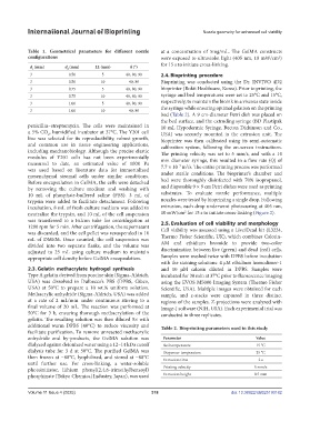
Table 1. Geometrical parameters for different nozzle at a concentration of 5mg/mL. The GelMA constructs
configurations were exposed to ultraviolet light (405 nm, 10 mW/cm )
2
d (mm) d (mm) LL (mm) θ (°) for 15 s to initiate cross-linking.
1
2
3 0.50 5 40, 80, 90 2.4. Bioprinting procedure
3 0.50 10 40, 80 Bioprinting was conducted using the Dr. INVIVO 4D2
3 0.75 5 40, 80, 90 bioprinter (Rokit Healthcare, Korea). Prior to printing, the
3 0.75 10 40, 80, 90 syringe and bed temperatures were set to 25°C and 15°C,
3 1.00 5 40, 80, 90 respectively, to maintain the bioink in a viscous state inside
3 1.00 10 40, 80 the syringe while ensuring optimal gelation on the printing
bed (Table 2). A 9 cm-diameter Petri dish was placed on
the bed surface, and the extruding syringe (BD Plastipak
penicillin–streptomycin. The cells were maintained in 10 mL Hypodermic Syringe, Becton Dickinson and Co.,
a 5% CO , humidified incubator at 37°C. The Y201 cell USA) was securely mounted in the extrusion unit. The
2
line was selected for its reproducibility, robust growth, bioprinter was then calibrated using its semi-automatic
and common use in tissue engineering applications, calibration system, following the on-screen instructions.
including mechanobiology. Although the precise elastic The printing velocity was set to 5 mm/s, and with a 14
modulus of Y201 cells has not been experimentally mm diameter syringe, this resulted in a flow rate (Q) of
measured to date, an estimated value of 1000 Pa −7 3
was used based on literature data for immortalized 7.7 × 10 m /s. The entire printing process was performed
mesenchymal stromal cells under similar conditions. under sterile conditions. The bioprinter’s chamber and
Before encapsulation in GelMA, the cells were detached bed were thoroughly disinfected with 70% isopropanol,
by removing the culture medium and washing with and disposable 9 × 9 cm Petri dishes were used as printing
10 mL of phosphate-buffered saline (PBS). 3 mL of substrates. To evaluate nozzle performance, multiple
trypsin were added to facilitate detachment. Following nozzles were tested by bioprinting a single drop. Following
incubation, 8 mL of fresh culture medium was added to extrusion, each drop underwent photocuring at 405 nm,
2
neutralize the trypsin, and 10 mL of the cell suspension 10 mW/cm for 15 s to initiate cross-linking (Figure 2).
was transferred to a Falcon tube for centrifugation at 2.5. Evaluation of cell viability and morphology
1200 rpm for 5 min. After centrifugation, the supernatant Cell viability was assessed using a Live/Dead kit (L3224,
was discarded, and the cell pellet was resuspended in 10
mL of DMEM. Once counted, the cell suspension was Thermo Fisher Scientific, UK), which combines Calcein-
divided into two separate flasks, and the volume was AM and ethidium bromide to provide two-color
adjusted to 25 mL using culture medium to maintain discrimination between live (green) and dead (red) cells.
appropriate cell density before GelMA encapsulation. Samples were washed twice with DPBS before incubation
with the staining solutions: 4 µM ethidium homodimer-1
2.3. Gelatin methacrylate hydrogel synthesis and 10 µM calcein diluted in DPBS. Samples were
Type A gelatin derived from porcine skin (Sigma-Aldrich, incubated for 30 min at 37°C prior to fluorescence imaging
USA) was dissolved in Dulbecco’s PBS (DPBS, Gibco, using the EVOS M5000 Imaging System (Thermo Fisher
USA) at 50°C to prepare a 10 wt.% uniform solution. Scientific, USA). Multiple images were obtained for each
Methacrylic anhydride (Sigma-Aldrich, USA) was added sample, and z-stacks were captured in three distinct
at a rate of 2 mL/min under continuous stirring to a regions of the samples. Z-projections were analyzed with
final volume of 20 mL. The reaction was performed at Image-J software (NIH, USA). Each experimental trial was
50°C for 3 h, ensuring thorough methacrylation of the conducted in three replicates.
gelatin. The resulting solution was then diluted 5× with
additional warm DPBS (40°C) to reduce viscosity and Table 2. Bioprinting parameters used in this study
facilitate purification. To remove unreacted methacrylic
anhydride and by-products, the GelMA solution was Parameter Value
dialyzed against deionized water using a 12–14 kDa cutoff Bed temperature 15 °C
dialysis tube for 3 d at 50°C. The purified GelMA was Dispenser temperature 25 °C
then frozen at −80°C, lyophilized, and stored at −80°C Extrusion time 2 s
until further use. For cross-linking, a water-soluble
photoinitiator, lithium phenyl(2,4,6-trimethylbenzoyl) Printing velocity 5 mm/s
phosphinate (Tokyo Chemical Industry, Japan), was used Extrusion height 0.5 mm
Volume 11 Issue 4 (2025) 318 doi: 10.36922/IJB025190182