
Page 354 - v11i4
P. 354
International Journal of Bioprinting GradGelMA 3D-bioprinted vascular skin
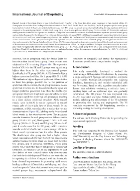
Figure 9. Repair of dorsal skin defects in New Zealand rabbits. (A) Flowchart of the dorsal skin defect repair experiment in New Zealand rabbits. (B)
Photographs of dorsal skin defect healing in New Zealand rabbits at Day 0, Day 7, Day 14, Day 21, and Day 28. By Day 28, all groups except the control group
had completely healed. At Day 21, the endothelial keratinocyte skin (EK) group had already achieved complete healing, showing significant differences
compared to the control group (96.43 ± 0.71%), blank group (98.16 ± 0.46%), and epidermis skin (E) group (99.48 ± 0.38%). (C) Hematoxylin and eosin
staining revealed that the EK and E groups had developed a “ridge-like” structure in the epidermis. However, the dermo-epidermal junction in the E group
was less defined compared to the EK group. The epidermal thickness in the EK group (56.59 ± 10.68 μm) was significantly greater than that in the E group
(45.65 ± 13.81 μm). Scale bars: 50 and 200 µm; magnification: 200× and 40×. (D) Cytokeratin 10 (CK10) protein expression in the newly formed epidermis
of New Zealand rabbits. The EK group showed significantly higher CK10 expression than the control and blank groups, but no significant difference was
observed compared to the E group. Scale bars: 20 and 100 µm. (E) Alpha-smooth muscle actin protein expression in small blood vessels and measurement
of the maximum vascular diameter in the newly formed skin of New Zealand rabbits. The EK group exhibited the largest vascular diameter (95.36 ± 24.87
μm), which was significantly different compared to the control group (13.53 ± 3.32 μm), blank group (17.69 ± 4.13 μm), and E group (35.83 ± 11.70 μm).
Scale bars: 50 and 100 µm. Data were analyzed via a one-way analysis of variance and are shown as mean ± standard deviation (*p < 0.05, **p < 0.01, and
***p < 0.001, n = 3). Abbreviation: IOD, integrated optical density.
structure, but its integration with the dermal layer was substitutes or autografts) and extend the experimental
less robust than that of the EK group. Tissue sections were duration to provide more comprehensive insights.
subjected to CK10 staining (Figure 9D). The expression
levels of CK10 in the EK and E groups were significantly 4. Conclusion
higher than those in the other experimental groups. This study successfully developed a method for
Specifically, the EK group (143.84 ± 8.55) showed a slightly constructing a 3D bioprinted VS substitute. By proposing
higher expression level than the E group (128.30 ± 4.83). a single-component hydrogel-cell-compatible composite
This indicates a higher degree of epidermal differentiation ink, a GelMA hydrogel-cell-compatible ink targeting
in these two groups, possibly due to the presence of fibroblasts, keratinocytes, and vascular endothelial cells
epidermal cells. In contrast, the EK group has a more mature was formulated. Based on this, a gradient vascularization
epidermal structure due to its accelerated early repair and dermal skin substitute containing a reticular layer, a
longer epidermal generation time than the double-layer papillary layer, and an epidermal layer was gradually
skin group. Moreover, it had more vascular cells/structures constructed. The 3D-printed VS was implanted into
for oxygen supply for epidermal cell generation, resulting BALB/c nude mice and New Zealand rabbit skin defect
in a more mature epidermal structure. Alpha-smooth models, validating the effectiveness of the VS substitute
muscle actin (α-SMA) is mainly expressed in smooth in promoting skin healing and angiogenesis. The VS
muscle cells of the middle layer of blood vessels. In this substitute constructed by 3D bioprinting provides a
experiment, α-SMA was selected as a marker for vascular promising strategy for treating skin injuries.
characterization (Figure 9E), labeling newly formed small
blood vessels in the subcutaneous tissue. The maximum Acknowledgments
vascular diameters for each group were as follows: control The authors thank Zhejiang University for providing the
group (13.53 ± 3.32 μm), Blank group (17.69 ± 4.13 μm), experimental equipment.
E group (35.83 ± 11.70 μm), and EK group (95.36 ± 24.87
μm). It can be seen that the EK group, which contained Funding
vascular endothelial cells, had a much stronger ability for
blood vessel regeneration than the other three groups. This work was supported by the National Key Research
and Development Program of China (Grant No.
The double-layer skin group also had a higher blood 2018YFA0703000) and the Key Science and Technology
vessel regeneration ability than the first two experimental Program of Zhejiang Province (Grant No. 2023C03170
samples because its repair speed was faster than the other and 2023C03071).
two groups, and it contained fibroblasts, which could
secrete VEGF and other factors that promote vascular cell Conflict of interest
formation. In summary, the 28-day experiment on back
skin defects of New Zealand rabbits fully demonstrated The authors declare no conflict of interest.
that both the double-layer and VS substitutes were effective
in repairing skin defects. Compared with the former, the Author contributions
latter showed improvements in promoting rapid wound Conceptualization: Yichen Luo, Bin Zhang, Jien Ma
healing, epidermal differentiation and shaping, and blood Data curation: Yichen Luo, Dan Li, Bin Zhang
vessel neogenesis. Future studies must incorporate positive Figure and visualization: Yichen Luo, Dan Li
control groups (e.g., commercial collagen-based skin Formal analysis: Yichen Luo
Volume 11 Issue 4 (2025) 346 doi: 10.36922/IJB025090069