
Page 364 - v11i4
P. 364
International Journal of Bioprinting Sr-doped printed scaffolds for bone repair
penetration of the cranial bone. A small number of the 1 min. The sections were then stained with hematoxylin
cranial connections were retained. After testing the skull staining solution for 3–5 min, washed with tap water,
with ophthalmic forceps for looseness, the skull was differentiated with differentiation solution, washed again
removed along the peripheral circular defect. The defect with tap water, returned to blue using the “Return to Blue
area was repeatedly rinsed with saline, and the scaffold was Solution,” and finally rinsed with running water. Later,
implanted into the cranial defect site of rats; no material the slices were dehydrated in 95% alcohol for 1 min, then
was placed in the blank group. The wound was closed using stained in an eosin staining solution for 15 s. The sections
a 4-0 absorbable suture, and antibiotics were injected for were sequentially dehydrated in 80% anhydrous ethanol
3 consecutive days postoperation to prevent infection. (2 min), 95% ethanol (2 min), and 100% ethanol (2 min),
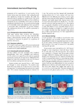
The implantation process of the scaffold is presented in followed by clearing in n-butanol (2 × 2 min) and xylene
Figure 2A–C. (2 × 2 min). Sections were then mounted using neutral
gum. Under a light microscope, the nuclei appeared blue
2.5.2. Postoperative observational indicators and the cytoplasm red.
Vital signs, activity, diet, urination and defecation,
and incision recovery of SD rats were observed after 2.5.5. Masson staining
surgery, with particular attention paid to the presence Cranial specimens with completed decalcification were
of abdominal distension to prevent the occurrence of fixed, embedded, and sectioned. Paraffin sections were
intestinal obstruction. deparaffinized in water by sequential immersion in xylene
for 20 min (twice), followed by 95% ethanol for 5 min,
2.5.3. Specimen collection 80% ethanol for 5 min, and 75% ethanol for 5 min, before
At 1, 2, and 3 months post-surgery, SD rats were euthanized washing with tap water.
to obtain liver, kidney, and skull specimens, which were
immersed in 4% paraformaldehyde. Frozen sections were removed from the –20°C freezer,
brought to room temperature, fixed with tissue fixative
2.5.4. Hematoxylin and eosin staining for 15 min, and rinsed with running water. The sections
Rat liver, kidney, and skull specimens with completed were immersed in Weigert’s iron hematoxylin solution
decalcification were fixed, embedded, and sectioned. overnight, followed by rinsing under running tap water.
Paraffin sections were deparaffinized in water by sequential Sections were immersed in a 1:1 mixture of Lichun Red
immersion in xylene for 20 min (twice), followed by 95% Acidic Compound Red solution (a mixed dyeing solution
ethanol for 5 min, 80% ethanol for 5 min, and 75% ethanol composed of the main dyes Acid Red 112 and Acid Red 87)
for 5 min, before washing them with tap water. and 1% phosphomolybdic acid aqueous solution for 1 min,
Frozen sections were removed from a –20°C freezer washed with tap water, differentiated for a few seconds in
and brought to room temperature. The sections were differentiation solution, and finally rinsed with tap water.
fixed with tissue fixative for 15 min and then rinsed with The slices were stained with aniline blue solution for
running water. For pretreatment, the sections were treated 6 min and then rinsed in tap water. They were subsequently
with “HD Constant Staining Pretreatment Solution” for immersed in 0.2% glacial acetic acid for 1 min. Without
Figure 2. Preparation of the bilateral cranial bone defect model (diameter: 5 mm) and implantation of scaffolds in SD rats. (A) Establishment of bilateral
cranial defects in SD rats. (B) Scaffolds implantation process. (C) Scaffolds implantation in the defect area. Abbreviation: SD, Sprague-Dawley.
Volume 11 Issue 4 (2025) 356 doi: 10.36922/IJB025210211