
Page 366 - v11i4
P. 366
International Journal of Bioprinting Sr-doped printed scaffolds for bone repair
Figure 3. Characterization of SrBG. (A) SEM image of the surface morphology of SrBG. (B) Particle size distribution of SrBG. Scale bar: 8 μm (A).
Abbreviations: SEM, scanning electron microscope; SrBG, strontium doped bioglass.
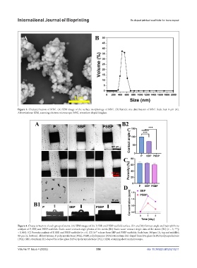
Figure 4. Characterization of each group of stents. (A) SEM images of the P, SBP, and PSBP scaffold surface. (B1 and B2) Contact angle and hydrophilicity
analysis of P, SBP, and PSBP scaffolds. Static water contact angle photos of the stents (B1) Static water contact Angle data of the stents (B2) (n = 3; ***p
< 0.001). (C) Porosity analysis of P, SBP, and PSBP scaffolds (n = 6). (D) Sr² release from SBP and PSBP scaffolds. Scale bars: 300 μm (A, top and middle);
+
80 μm (A, bottom). Abbreviations: P, polycaprolactone (PCL); PSBP, polydopamine (PDA)/strontium (Sr)-doped bioactive glass (SrBG)/polycaprolactone
(PCL); SBP, strontium (Sr)-doped bioactive glass (SrBG)/polycaprolactone (PCL); SEM, scanning electron microscope.
Volume 11 Issue 4 (2025) 358 doi: 10.36922/IJB025210211