
Page 371 - v11i4
P. 371
International Journal of Bioprinting Sr-doped printed scaffolds for bone repair
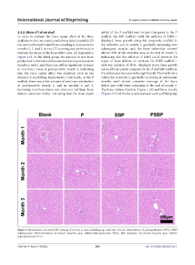
3.3.2. Micro-CT of rat skull ability of the P scaffold was limited. Compared to the P
In order to evaluate the bone repair effect of the three scaffold, the SBP scaffold—with the addition of SrBG—
scaffolds in vivo, we constructed a bone defect model in SD displayed bone growth along the composite scaffold in
rats and performed cranial bone sampling at postoperative the defective area at month 1, gradually increasing over
months 1, 2, and 3; micro-CT scanning was performed to subsequent months until the bone trabeculae covered
evaluate the repair of the bone defect area. As displayed in almost 50% of the defective area at the end of month 3,
Figure 11A, in the blank group, the amount of new bone indicating that the addition of SrBG could promote the
production in the bone defect area was low at postoperative repair of bone defects. In contrast, the PSBP scaffold—
months 1 and 2, and there was still no significant increase with the addition of PDA—displayed more bone growth
in new bone tissue at postoperative month 3, indicating across all time points compared to the P and SBP scaffolds;
that the bone repair effect was relatively poor in the the defect area was seen to be significantly filled with bone
absence of scaffolding implantation. Conversely, in the P trabeculae at month 1, gradually increasing in subsequent
scaffold, there was a low amount of new bone production months until almost complete coverage of the bone
at postoperative month 1, and in months 2 and 3, defect area with bone trabeculae at the end of month 3.
increasing new bone tissue was observed, but large bone The bone volume fraction (Figure 11B) and bone density
defects remained visible, indicating that the bone repair (Figure 11C) of the bone defect area in each scaffold group
Figure 9. Hematoxylin and eosin (HE) staining of rat liver in each scaffold group. Scale bar: 100 μm. Abbreviations: P, polycaprolactone (PCL); PSBP,
polydopamine (PDA)/strontium (Sr)-doped bioactive glass (SrBG)/polycaprolactone (PCL); SBP, strontium (Sr)-doped bioactive glass (SrBG)/
polycaprolactone (PCL).
Volume 11 Issue 4 (2025) 363 doi: 10.36922/IJB025210211