
Page 367 - v11i4
P. 367
International Journal of Bioprinting Sr-doped printed scaffolds for bone repair
continuously released Sr² over time, but the total Sr² (Figure 5B2). With the gradual addition of SrBG and PDA,
+
+
released from the PSBP scaffolds was lower than that of the the compressive strength and compressive modulus of the
SBP scaffolds (Figure 4D). P, SBP, and PSBP scaffolds gradually increased, and the
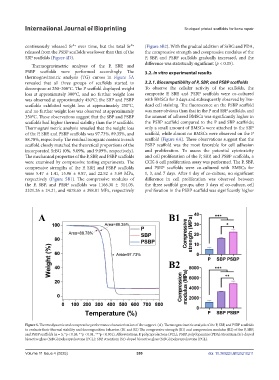
Thermogravimetric analyses of the P, SBP, and difference was statistically significant (p < 0.05).
PSBP scaffolds were performed accordingly. The 3.2. In vitro experimental results
thermogravimetric analysis (TG) curves in Figure 5A
revealed that all three groups of scaffolds started to 3.2.1. Biocompatibility of P, SBP, and PSBP scaffolds
decompose at 250–300°C. The P scaffold displayed weight To observe the cellular activity of the scaffolds, the
loss at approximately 300°C, and no further weight loss composite P, SBP, and PSBP scaffolds were co-cultured
was observed at approximately 450°C; the SBP and PSBP with BMSCs for 3 days and subsequently observed by live-
scaffolds exhibited weight loss at approximately 250°C, dead cell staining. The fluorescence on the PSBP scaffold
and no further weight loss was observed at approximately was more obvious than that in the P and SBP scaffolds, and
350°C. These observations suggest that the SBP and PSBP the amount of adhered BMSCs was significantly higher in
scaffolds had higher thermal stability than the P scaffolds. the PSBP scaffold compared to the P and SBP scaffolds;
Thermogravimetric analysis revealed that the weight loss only a small amount of BMSCs were attached to the SBP
of the P, SBP, and PSBP scaffolds was 97.73%, 89.35%, and scaffold, while almost no BMSCs were observed on the P
88.78%, respectively. The residual inorganic content in each scaffold (Figure 6A). These observations suggest that the
scaffold closely matched the theoretical proportions of the PSBP scaffold was the most favorable for cell adhesion
incorporated SrBG (0%, 9.09%, and 9.09%, respectively). and proliferation. To assess the potential cytotoxicity
The mechanical properties of the P, SBP, and PSBP scaffolds and cell proliferation of the P, SBP, and PSBP scaffolds, a
were examined by compressive testing experiments. The CCK-8 cell proliferation assay was performed. The P, SBP,
compressive strengths of the P, SBP, and PSBP scaffolds and PSBP scaffolds were co-cultured with BMSCs for
were 5.47 ± 1.41, 15.96 ± 0.87, and 22.52 ± 3.69 MPa, 1, 3, and 7 days. After 1 day of co-culture, no significant
respectively (Figure 5B1). The compressive modulus of difference in cell proliferation was observed between
the P, SBP, and PSBP scaffolds was 1166.30 ± 314.05, the three scaffold groups; after 3 days of co-culture, cell
3321.26 ± 24.21, and 4878.65 ± 398.81 MPa, respectively proliferation in the PSBP scaffold was significantly higher
Figure 5. Thermodynamic and compressive performance characterization of the support. (A). Thermogravimetric analysis of the P, SBP, and PSBP scaffolds
to evaluate their thermal stability and decomposition behavior. (B1 and B2) The compressive strength (B1) and compression modulus (B2) of the P, SBP,
and PSBP scaffolds (n = 3; *p < 0.05, **p < 0.01, ***p < 0.001). Abbreviations: P, polycaprolactone (PCL); PSBP, polydopamine (PDA)/strontium (Sr)-doped
bioactive glass (SrBG)/polycaprolactone (PCL); SBP, strontium (Sr)-doped bioactive glass (SrBG)/polycaprolactone (PCL).
Volume 11 Issue 4 (2025) 359 doi: 10.36922/IJB025210211