
Page 372 - v11i4
P. 372
International Journal of Bioprinting Sr-doped printed scaffolds for bone repair
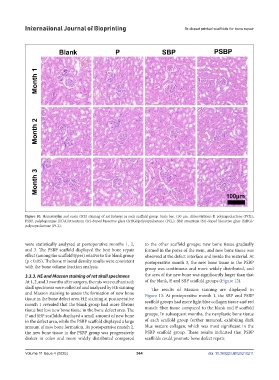
Figure 10. Hematoxylin and eosin (HE) staining of rat kidneys in each scaffold group. Scale bar: 100 μm. Abbreviations: P, polycaprolactone (PCL);
PSBP, polydopamine (PDA)/strontium (Sr)-doped bioactive glass (SrBG)/polycaprolactone (PCL); SBP, strontium (Sr)-doped bioactive glass (SrBG)/
polycaprolactone (PCL).
were statistically analyzed at postoperative months 1, 2, to the other scaffold groups; new bone tissue gradually
and 3. The PSBP scaffold displayed the best bone repair formed in the pores of the stent, and new bone tissue was
effect (among the scaffold types) relative to the blank group observed at the defect interface and inside the material. At
(p < 0.05). The bone mineral density results were consistent postoperative month 3, the new bone tissue in the PSBP
with the bone volume fraction analysis. group was continuous and more widely distributed, and
3.3.3. HE and Masson staining of rat skull specimens the area of the new bone was significantly larger than that
At 1, 2, and 3 months after surgery, the rats were euthanized; of the blank, P, and SBP scaffold groups (Figure 12).
skull specimens were collected and analyzed by HE staining The results of Masson staining are displayed in
and Masson staining to assess the formation of new bone Figure 13. At postoperative month 1, the SBP and PSBP
tissue in the bone defect area. HE staining at postoperative scaffold groups had more light blue collagen tissue and red
month 1 revealed that the blank group had more fibrous
tissue but less new bone tissue in the bone defect area. The muscle fiber tissue compared to the blank and P scaffold
P and SBP scaffolds displayed a small amount of new bone groups. In subsequent months, the neoplastic bone tissue
in the defect area, while the PSBP scaffold displayed a large of each scaffold group further matured, exhibiting dark
amount of new bone formation. At postoperative month 2, blue mature collagen, which was most significant in the
the new bone tissue in the PSBP group was progressively PSBP scaffold group. These results indicated that PSBP
darker in color and more widely distributed compared scaffolds could promote bone defect repair.
Volume 11 Issue 4 (2025) 364 doi: 10.36922/IJB025210211