
Page 370 - v11i4
P. 370
International Journal of Bioprinting Sr-doped printed scaffolds for bone repair
3.3. In vivo experimental results in these organs. It was observed that specific structures
in the rat liver (Figure 9) and kidney (Figure 10)—such
3.3.1. Hepatotoxicity and nephrotoxicity of P, SBP, and as the central vein, the confluent area, hepatocytes,
PSBP scaffolds glomeruli, and mesangial cells—in each scaffold group
To assess the biosafety of the scaffolds, liver and kidney had a clear morphology and boundaries, with no signs
tissues of rats in each scaffold group were sampled at of inflammatory cell infiltration. This suggests that the
postoperative months 1, 2, and 3, and tissue sections were composite scaffolds have good biocompatibility, as they
HE-stained to observe the presence or absence of lesions did not have any toxicity effects on the rat liver and kidney.
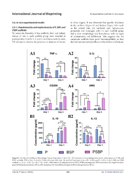
Figure 8. The effect of scaffolds on Macrophage Typing Polarization in vitro. (A1–A4) Expression of macrophage polarization-related genes on P, SBP, and
PSBP scaffolds: TNF-α (A1), IL1β (A2), CD206 (A3), and ARG (A4). (B1 and B2) Expression levels of IL-10 (B2) and IL-12 (B1) in the P, SBP, and PSBP
scaffolds. n = 3; *p < 0.05, **p < 0.01, ***p < 0.001. Abbreviations: P, polycaprolactone (PCL); PSBP, polydopamine (PDA)/strontium (Sr)-doped bioactive
glass (SrBG)/polycaprolactone (PCL); SBP, strontium (Sr)-doped bioactive glass (SrBG)/polycaprolactone (PCL).
Volume 11 Issue 4 (2025) 362 doi: 10.36922/IJB025210211