
Page 65 - JCTR-11-1
P. 65
Journal of Clinical and
Translational Research Osteoporosis risk factors in diabetics
significant difference in BMI between the osteoporosis
group with osteopenia and normal DEXA in both sexes.
The differences in WHR were inconsistent and did not
exhibit a trend in the male and female groups.
The anthropometric and biochemical parameters
usually advised during the follow-up of T2DM patients
were used as the dependent variables to predict the T-score
of the lumbar vertebra in all patients (Tables 5 and 6).
The overall multiple linear regression was statistically
significant (R = 0.307; F = 3.402; regression degrees
2
of freedom [df regression ] = 6; residual degrees of freedom
[df residual ] = 196; P = 0.003). The T-score was obtained using
a constant of −9.531 and the respective values of BMI
(0.139), WHR (0.185), age (0.081), duration of T2DM
(−0.068), HbA1c (0.053), urine ACR (0.06), and duration
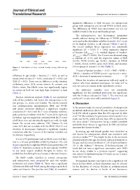
Figure 2. Distribution of bone mineral density among different age of menopause in women (−0.186) (Table 7):
groups T-score of lumbar vertebra = −9.531 + BMI + WHR +
HbA1c + duration of T2DM (years) + age (years) + urine
difference in age (males > females; P < 0.01), as well as ACR + duration of menopause in women (I)
serum levels of urea (P = 0.01), creatinine (P < 0.01), and
TSH (P = 0.02). There was no difference in the duration Where the duration of menopause will only apply to
of diabetes, urine ACR, serum vitamin D, calcium, and women who have attained menopause; its value will be
HbA1c levels. The FRAX score was significantly higher zero for men and women in the reproductive period.
in women in both left and right hips compared to men The individual variables were not statistically
(Table 1). significant, but the combined prediction was significant,
with the P-values indicated in Table 7. The observed and
Pearson correlation analysis (Table 2) was performed predicted T-scores were well-correlated (Figure 3).
with all the probable risk factors for osteoporosis in the
two groups, i.e., males and females. The results revealed 4. Discussion
that anthropometric measurements (BMI and WHR)
and serum creatinine displayed a significant negative In the present study, the overall prevalence of osteoporosis
correlation (P < 0.01) with the T-score of lumbar vertebrae in diabetic individuals above 50 years of age was found to
be 40.9%, which is greater than the 33% reported by Aleti
and a positive correlation with the T-score of the right and et al. On the contrary, the prevalence of osteopenia in our
14
left femur. Age was negatively correlated with the T-scores study was 32.5%, which is lower than 40% as reported by
of all three sites and statistically significant with the right the same study. Osteoporosis being a widely prevalent
14
femur. The serum urea, calcium, TSH, Vitamin D, and metabolic bone disease is aggravated in DM and further
urine ACR did not correlate with the BMD. HbA1C and increases the disease burden. 15
duration of menopause displayed a significant negative
correlation with the T-scores of the lumbar vertebra and Increasing age and female gender were identified as
the femurs, respectively. risk factors for osteoporosis, which was consistent with
previous studies. 14,16-18 This might be due to the decreased
A total of 53 participants had an FRAX score (left hip), physical activity with age, decreased calcium absorption in
that is, a 10-year probability of left hip fracture >3%, and the gut, and decreased synthesis of 1,25-dihydroxyvitamin
41 participants had a right hip FRAX score greater than 3% D3 in the kidneys. However, 25-hydroxyvitamin D3
19
(Table 3). Therefore, as many as 83 participants (40.89%) levels did not have a significant association with BMD
in our study require medical therapies to reduce the in our study, similar to a previous report, suggesting
11
probability of suffering from a fracture in the next 10 years. that altered 25-hydroxyvitamin D3 levels are not a major
The BMD results, classified as normal, osteopenia, reason for bone loss.
and osteoporosis, were compared for each risk variable Menopausal duration was found to be a significant risk
in females and males (Table 4). There was a significant factor for low BMD, as bone absorption may be affected
difference in BMI and WHR among the subgroups in both by the depletion of ovarian follicles in postmenopausal
males and females. The post hoc test (Table 4) revealed a women and the decline in levels of sex hormones.
Volume 11 Issue 1 (2025) 59 doi: 10.36922/jctr.24.00062