
Page 52 - JCTR-9-6
P. 52
416 Mustahsan et al. | Journal of Clinical and Translational Research 2023; 9(6): 414-422
randomized into two experimental groups (n = 7): (1) Untreated sterilized in 70% ethanol and implanted into the muscle pouch,
scaffolds and (2) decellularized scaffolds. Decellularized scaffolds and the fascia over the muscle was sutured with resorbable sutures
were prepared using the protocol explained in section 2.1. A set of (Ethicon, Raritan, NJ, USA) to close the muscle wound. The skin
untreated scaffolds which were exposed to the same reagents as incision was closed using non-resorbable sutures (Ethicon), and
decellularized scaffolds were also prepared, except that they do topical antibiotics were applied. Buprenorphine (0.1 mg/kg)
not have any stem cells seeded onto the surface. analgesia (Buprenex, Indivior, North Chesterfield, VA, USA)
was administered immediately following surgery and twice daily
2.3. Surgical protocol
thereafter until it was judged to be no longer necessary. The skin
For implantation, animals were anesthetized with isoflurane sutures were removed 10 – 14 days after surgery. The workflow of
(2% induction and 1.5% maintenance) (Covetrus, Portland, ME, the surgery is illustrated in Figure 2.
USA). Eye lubrication (Optixcare; Aventix, Burlington, Ontario, The animals were monitored every day for the first 3 days and
Canada) was applied, and the mice were prepared for the surgery weekly thereafter. Movement around the cage and activity was
by shaving the left hind limb and sterilizing the surgical site with observed to assess the weight-bearing on the lower extremity.
betadine and ethanol (Sigma-Aldrich, Saint-Louis, MO, USA). The incision area was assessed for incision and quality of sutures.
A 10-mm longitudinal incision parallel to the posterior femur Grooming, vocalization, and weight loss were checked as
was created. Using a blunt dissection to prevent muscle damage, indicators of distress. The exclusion criteria were if the animal is
a 5-mm deep intramuscular pouch was then shaped, taking experiencing dehiscence, infection, pain, or distress that cannot be
precautions not to expose the periosteum. A scaffold was, then, treated or if the animal is experiencing more than 15% weight loss.
A B C D
Figure 1. (A) The cross-section of a mouse vertebra with cortical bone (yellow) and trabecular bone (brown); (B) Trabecular bone extracted from
µ-computed tomography scan of the vertebra; (C) MED610 scaffold designed from the extracted trabecular bone seeded with bone cells on its surface;
and (D) Scaffold being implanted into the thigh muscle of a mouse.
A B C
D E F
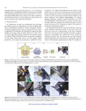
Figure 2. (A) Preparing the surgical site by scrubbing the shaved skin with Betadine: (B) Longitudinal incision made along the thigh; (C) Blunt
dissection being carried out to create a muscle pouch without exposing the periosteum; (D) Implant placed in the muscle pouch; (E) muscle pouch was
closed with resorbable sutures; and (F) The skin incision was sutured with non-resorbable sutures.
DOI: http://dx.doi.org/10.18053/jctres.09.202306.23-00097