
Page 51 - JCTR-9-6
P. 51
Mustahsan et al. | Journal of Clinical and Translational Research 2023; 9(6): 414-422 415
cost and donor site complications, have a higher chance of antigen assess the biocompatibility of the 3D-printed MED610 scaffold,
response and disease transmissibility [8-10]. Moreover, allografts seeded with MC3T3 stem cells, and treated with CRFP.
are weaker in comparison to metallic implants leading to fractures
and future revision surgeries [11,12]. 2. Materials and Methods
Synthetic bone graft substitutes (BGSs) are being developed 2.1. Preparation of MED610 scaffolds
to overcome limitations in conventional metallic and biological
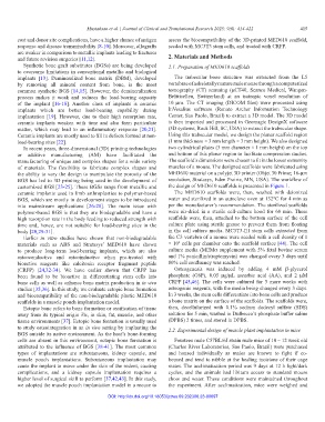
implants [13]. Demineralized bone matrix (DBM), developed The trabecular bone structure was extracted from the L5
by removing all mineral content from bone, is the most vertebrae of a skeletally mature male mouse through a computerized
common synthetic BGS [14,15]. However, the demineralization tomography (CT) scanning (µCT40, Scanco Medical, Wangen-
process makes it weak and reduces the load-bearing capacity Brüttisellen, Switzerland) at an isotropic voxel resolution of
of the implant [16-18]. Another class of implants is ceramic 10 µm. The CT imaging (DICOM files) were processed using
implants which are better load-bearing capability during InVesalius software (Renato Archer Information Technology
implantation [19]. However, due to their high resorption rate, Center, Sao Paolo, Brazil) to extract a 3D model. The 3D model
ceramic implants weaken with time and also form particulate is then imported and processed in Geomagic DesignX software
matter, which may lead to an inflammatory response [20,21]. (3D systems, Rock Hill, SC, USA) to extract the trabecular shape.
Ceramic implants are mostly used to fill in defects formed at non- Using this trabecular model, we design the planar scaffold region
load-bearing sites [22]. (1 mm thickness × 3 mm length × 3 mm height). We also designed
In recent years, three-dimensional (3D) printing technologies two cylindrical plates (3 mm diameter × 1 mm height) on the top
or additive manufacturing (AM) have facilitated the and bottom of this planar region to facilitate compression studies.
manufacturing of unique and complex shapes for a wide variety The scaffold’s dimensions were chosen to fit in the lower extremity
of materials. The flexibility to fabricate complex shapes and muscles of a mouse. The designed scaffolds were fabricated using
the ability to vary the design to manipulate the porosity of the MED610 material on a polyjet 3D printer (Objet 30 Prime; 16-µm
BGS has led to 3D printing being used in the development of resolution, Stratasys, Eden Prairie, MN, USA). The workflow of
customized BGS [23-25]. These BGSs range from metallic and the design of MED610 scaffolds is presented in Figure 1.
ceramic implants used in limb arthroplasties to polymer-based The MED610 scaffolds were, then, washed with deionized
BGS, which are mostly in development stages to be introduced water and sterilized in an autoclave oven at 132°C for 4 min as
into mainstream applications [26-28]. The main issue with per the manufacturer’s recommendation. The sterilized scaffolds
polymer-based BGS is that they are biodegradable and have a were air-dried in a sterile cell-culture hood for 60 min. These
high resorption rate in the body leading to reduced strength with scaffolds were, then, attached to the bottom surface of the cell
time and, hence, are not suitable for load-bearing sites in the culture plate using sterile grease to prevent them from floating
body [20,29-31]. in the cell culture media. MC3T3-E1 stem cells extracted from
Earlier in vitro studies have shown that non-biodegradable the C3 vertebra of a mouse were seeded with a cell density of 1
3
materials such as ABS and Stratasys’ MED610 have shown × 10 cells per chamber onto the scaffold surface [44]. The cell
to produce long-term load-bearing implants, which are also culture media (MEMα supplement with 5% fetal bovine serum
osteoconductive and osteoinductive when pre-treated with and 1% penicillin/streptomycin) was changed every 3 days until
bioactive reagents like calcitonin receptor fragment peptide 80% cell confluency was reached.
(CRFP) [24,32-34]. We have earlier shown that CRFP has Osteogenesis was induced by adding 4 mM β-glycerol
been found to be bioactive in differentiating stem cells into phosphate (G6P), 0.05 µg/µL ascorbic acid (AA), and 2 µM
bone cells as well as enhance bone matrix production in in vivo CRFP [45,46]. The cells were cultured for 3 more weeks with
studies [35,36]. In this study, we evaluate ectopic bone formation osteogenic reagents, with the media being changed every 3 days.
and biocompatibility of the non-biodegradable plastic MED610 In 3 weeks, the stem cells differentiate into bone cells and produce
scaffolds in a muscle pouch implantation model. a bone matrix on the surface of the scaffolds. The scaffolds were,
Ectopic bone refers to bone formation or ossification of tissue then, decellularized with 0.1% sodium dodecyl sulfate (SDS)
away from its typical origin Vis, in skin, fat, muscle, and other solution for 5 min, washed in Dulbecco’s phosphate buffer saline
tissue environments [37]. Ectopic bone formation is usually used (DPBS) 2 times, and stored in DPBS.
to study osteointegration in an in vivo setting by implanting the 2.2. Experimental design of muscle plant implantation in mice
BGS outside its native environment. As the host’s bone-forming
cells are absent in this environment, ectopic bone formation is Fourteen male C57BL/6J strain male mice of 10 – 12 week old
attributed to the influence of BGS [38-41]. The most common (Charles River Laboratories, Sao Paolo, Brazil) were purchased
types of implantations are subcutaneous, kidney capsule, and and housed individually as males are known to fight if co-
muscle pouch implantations. Subcutaneous implantation may housed and tend to nibble at the healing incisions of their cage
cause the implant to move under the skin of the rodent, causing mates. The acclimatization period was 9 days at 12 h light/dark
complications, and a kidney capsule implantation requires a cycles, and the animals had libitum access to standard mouse
higher level of surgical skill to perform [37,42,43]. In this study, chow and water. These conditions were maintained throughout
we adopted the muscle pouch implantation model in a mouse to the experiment. After acclimatization, mice were weighed and
DOI: http://dx.doi.org/10.18053/jctres.09.202306.23-00097