
Page 121 - MI-1-1
P. 121
Microbes & Immunity Rare multi-site Klebsiella pneumoniae infections
Laboratory testing revealed neutrophilic leukocytosis, respiratory failure, and metabolic acidosis. Laboratory
elevated levels of CRP (268 mg/L), PCT (15 ng/mL), testing revealed a WBC of 13.1×10 /L, with neutrophils
9
fasting BG (45 mmol/L), abnormal blood ketone bodies, comprising 96% of the total count. BG level was 33 mmol/L
and metabolic acidosis. ABG analysis while on 6 L/min and blood ketones tested positive. PCT was >10 ng/mL,
oxygen through nasal cannula demonstrated a pH of 7.0, while CRP was 389 mg/L. Imaging examinations, including
PaCO of 12 mmHg, PaO of 102 mmHg, HCO of 1.4 an abdominal CT scan and chest X-ray, revealed a sizable
-
3
2
2
mmol/L, lactic acid concentration of 8.7 mmol/L, and lesion in the right lobe of the liver, characterized by the
serum albumin level of 19 g/L. The patient underwent presence of gas and necrotic tissue (Figure 5). In response
intubation. X-ray examination revealed soft-tissue edema to the patient’s unstable hemodynamics, he was intubated
in the left leg, while abdominal CT scan findings revealed and administered vasopressors. Treatment was initiated
edema and gas accumulation along the left abdominal wall, with tigecycline 50 mg every 12 h and meropenem 1 g
as well as fluid retention in the left pelvis (Figure 4). Based every 8 h. At the same time, drainage of the liver abscess
on these findings, a diagnosis of necrotizing fasciitis, septic was performed. KP was identified in blood and pus sample
shock, and diabetic ketoacidosis was established. Empirical cultures and exhibited susceptibility to all tested antibiotics.
antibiotic treatment consisting of meropenem 1 g every Despite intensive medical care, the patient succumbed to
8 h and linezolid 0.6 g every 12 h was initiated. A surgical the serious infection 3 days after admission to the hospital.
decompression incision was performed on the left leg to
evacuate pus. However, despite intensive post-operative 2.6. Case 6
care, the patient succumbed to multiple organ failure A 72-year-old diabetic man presented to our hospital
6 days following hospital admission. KP susceptible to all with symptoms of chills, fever, left eye discomfort, and
tested antibiotics was identified in blood and infectious impaired vision persisting for a week. Notably, he had not
fluid cultures 3 days before the patient’s death. sought treatment for either his diabetes or hypertension.
2.5. Case 5 Laboratory testing revealed elevated levels of CRP
9
(219 mg/L) and WBC (21.9×10 /L). BG was 29 mmol/L,
A 61-year-old diabetic man presented to our hospital and blood ketones tested positive. Examination of the
with a history of fever, nausea, vomiting, and hepatic left eye revealed the presence of a hypopyon, which was
pain that had been ongoing for a week. These symptoms blocking the view of the retina and clouding the vitreous.
worsened over the course of a day. On assessment, his vital Brain magnetic resonance imaging revealed an abscess in
signs were notable for a temperature of 40.0°C, a heart the right temporal lobe of the brain and endophthalmitis
rate of 134 beats/min, a blood pressure of 54/34 mmHg, in the left eye. An abdominal CT scan identified a liver
a respiratory rate of 36 breaths/min, and an oxygen abscess (Figure 6). Based on these findings, a diagnosis
saturation of 85%. The patient had a longstanding history of endophthalmitis, liver abscess, brain abscess, diabetes
of untreated diabetes mellitus spanning 15 years. On arrival mellitus, and hypertension was established. Treatment was
at our hospital, he exhibited symptoms of septic shock, initiated with meropenem 1 g every 8 h and vancomycin
1 g every 12 h. KP was identified in the eye secretion sample
A C
culture and exhibited susceptibility to all tested antibiotics.
During the 5-day hospital stay, the patient reported
severe pain in his left eye. Subsequently, it was confirmed
that he had experienced lifelong visual loss, leading
to the decision to surgically remove the left eyeball.
A B
B
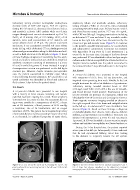
Figure 4. CT scan (A and B) and X-ray (C) findings for Case 4. The Figure 5. The liver abscess observed on medical imaging for Case 5.
patient suffered from necrotizing fasciitis in the left lower limb and left (A) A large liver abscess visualized on the abdominal computed
side of the hip. tomography scan. (B) The liver abscess depicted on the chest X-ray.
Volume 1 Issue 1 (2024) 115 doi: 10.36922/mi.2600