
Page 120 - MI-1-1
P. 120
Microbes & Immunity Rare multi-site Klebsiella pneumoniae infections
2.2. Case 2 12.1 mmol/L, base excess of −23.5 mmol/L, and lactic
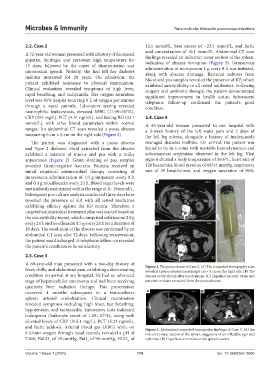
A 72-year-old woman presented with a history of decreased acid concentration of 10.1 mmol/L. Abdominal CT scan
appetite, lumbago, and persistent high temperature for findings revealed an indistinct outer section of the spleen,
15 days, followed by the onset of disorientation and indicative of abscess formation (Figure 3). Intravenous
administration of meropenem 1 g every 8 h was initiated,
nonsensical speech. Notably, she had left her diabetes along with abscess drainage. Bacterial cultures from
mellitus untreated for 20 years. On admission, the blood and pus samples revealed the presence of KP, which
patient exhibited resistance to physical examination. exhibited susceptibility to all tested antibiotics. Following
Clinical evaluation revealed symptoms of high fever, surgery and antibiotic therapy, the patient demonstrated
rapid breathing, and tachycardia. Her oxygen saturation significant improvement in health status. Subsequent
level was 91% despite receiving 6 L of oxygen per minute telephone follow-up confirmed the patient’s good
through a nasal cannula. Laboratory testing revealed condition.
9
neutrophilic leukocytosis, elevated WBC (17.09×10 /L),
CRP (255 mg/L), PCT (4.31 ng/mL), and fasting BG (33.1 2.4. Case 4
mmol/L), with other blood parameters within normal A 45-year-old woman presented to our hospital with
ranges. An abdominal CT scan revealed a psoas abscess a 3-week history of the left waist pain and 2 days of
measuring 6 cm × 8 cm on the right side (Figure 2). the left leg edema, alongside a history of inadequately
The patient was diagnosed with a psoas abscess managed diabetes mellitus. On arrival, the patient was
and Type 2 diabetes. Fluid extracted from the abscess found to be in a coma with unstable hemodynamics and
exhibited a mixture of mucus and pus with a milky subcutaneous crepitation observed in the left leg. Vital
appearance (Figure 2). Gram staining of pus samples signs indicated a body temperature of 39.6°C, heart rate of
revealed Gram-negative bacteria. Patients received an 126 beats/min, blood pressure of 68/44 mmHg, respiratory
initial empirical antimicrobial therapy consisting of rate of 29 breaths/min, and oxygen saturation of 90%.
intravenous administration of 1.0 g imipenem every 8 h
and 0.4 g moxifloxacin every 24 h. Blood sugar levels were A C
meticulously maintained within the range of 8– 10 mmol/L.
Subsequent pus culture analysis conducted three days later
revealed the presence of KP, with all tested medicines
exhibiting efficacy against the KP strains. Therefore, a
targeted antimicrobial treatment plan was started based on
the susceptibility report, which comprised ceftriaxone 2.0 g
every 24 h and levofloxacin 0.5 g every 24 h for a duration of B
10 days. The resolution of the abscess was confirmed by an
abdominal CT scan after 32 days. Following recuperation,
the patient was discharged. A telephone follow-up revealed
the patient’s condition to be satisfactory.
2.3. Case 3
A 69-year-old man presented with a two-day history of Figure 2. The psoas abcess in Case 2. (A) The computed tomography scan
fever, chills, and abdominal pain, exhibiting a deteriorating revealed a psoas abscess measuring 6 cm × 8 cm on the right side. (B) The
condition on arrival at our hospital. He had an advanced abscess cavity shrank after pus drainage. (C) Liquefied necrotic tissue and
stage of hepatocellular carcinoma and had been receiving purulent exudate extracted from the psoas abscess.
quarterly liver radiation therapy. This presentation
occurred 4 months subsequent to a transcatheter A B
splenic arterial embolization. Clinical examination
revealed symptoms including high fever, fast breathing,
hypotension, and tachycardia. Laboratory tests indicated
leukopenia (leukocyte count of 1.09×10 /L), along with
9
elevated levels of CRP (315.1 mg/L), PCT (5.23 ng/mL),
and lactic acidosis. Arterial blood gas (ABG) while on Figure 3. Abdominal computed tomography findings of Case 3. (A) An
6 L/min oxygen through nasal cannula revealed a pH of indistinct outer section of the spleen, suggestive of an inflatable sign and
7.010, PaCO of 18 mmHg, PaO of 96 mmHg, HCO of ischemia. (B) Liquefactive necrosis in the spleen’s cortex.
-
3
2
2
Volume 1 Issue 1 (2024) 114 doi: 10.36922/mi.2600