
Page 104 - MSAM-4-1
P. 104
Materials Science in Additive Manufacturing Topology optimization of an aluminum bicycle pedal
crank using laser powder bed fusion
dispersive X-ray spectroscopy electron microscopy at 1 µm cloth, and etching (0.5% hydrofluoric acid + 99.5%
the Materials Centre of the University of Porto (CEMUP, water) for 1 min. Micrography was obtained using the
Porto, Portugal). Leica DVM6 (Wetzlar, Germany) equipment and its
®
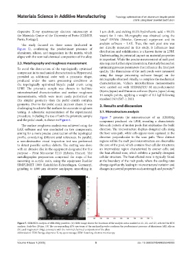
The study focused on three zones (indicated in analysis software – LAX. Thus, the melt pool size was
Figure 5), confirming the predominant presence of not directly measured in this study, it influences heat
aluminum, silicon, and magnesium in the matrix, which distribution and solidification is a known factor in LPBF.
aligns with the nominal chemical composition of the alloy. Understanding its potential impact on material properties
is important. While the precise measurement of melt pool
2.3. Metallography and roughness measurement size requires further experimentation, this study focused on
To avoid the destruction of the topologically optimized optimizing process parameters that influence the final part
component in its mechanical characterization, Hypermetal quality. The dimensions of the melt pools were measured
provided an additional cube with a prismatic shape, using the image processing software ImageJ on the
produced under the same processing conditions as micrographs obtained. Finally, to complete the mechanical
the topologically optimized bicycle pedal crank using characterization, Vickers microhardness measurements
LPBF. The prismatic sample was chosen to facilitate were carried out with SHIMADZU M microdurometer
microstructural characterization and surface roughness (Kyoto, Japan) and Duramin software (Kyoto, Japan) along
measurements, which were more easily performed on 14 sample points, applying a weight of 0.3 kgf following
this simpler geometry than the pedal crank’s complex standard ISO 6507-1: 2011.
geometry. Due to the pedal crank intricate shape, it was 3. Results and discussion
challenging to achieve flat surfaces for accurate roughness
testing. A schematic representation of the experimental 3.1. Microstructure analysis
procedure, including the use of both the prismatic sample Figure 7 presents the microstructure of an AlSi10Mg
and the pedal crank, is shown in Figure 6. component produced via LPBF, revealing a characteristic
The surface roughness analysis was achieved using the fish-scale pattern of molten pools that emphasizes the build
LAX software and was conducted on two components, direction. The microstructure displays elongated cells along
aiming for a more precise construction of the topological the laser scan path, while cells appear more equiaxed in the
profile, considering different axial references. In parallel, direction perpendicular to the scan path. Three distinct
a non-destructive visual inspection test was performed regions within the melt pool microstructure are observable:
to detect possible surface defects. The cutting was done the core of the pool, which contains finer cellular structures;
with an abrasive disc in the equipment designated for this an intermediate region characterized by coarser cells; and
purpose – Presi Mecatome T210 (Eybens, France). The the heat-affected zone, which exhibits a partially disrupted
metallographic preparation comprised the steps of hot cellular structure. The heat-affected zone is typically found
mounting in acrylic resin, using the equipment Buehler at the boundary of the melt pools, where the cooling rates
SIMPLIMET 1000 (Leinfelden-Echterdingen, Germany), diverge significantly, leading to microstructural variation and
grinding to 4000 µm abrasive sandpaper, smoothing in changes in material properties such as strength and porosity. 38
A B
Figure 5. SEM/EDS analysis of AlSi10Mg powders. (A) SEM image shows the locations of the analysis areas marked as Z1, Z2, and Z3, selected for EDS
analysis. Scale bar: 20 µm. (B) The corresponding EDS spectrum of the analyzed powder confirms the predominant presence of aluminum (Al), silicon
(Si), and magnesium (Mg), consistent with the nominal chemical composition of the alloy
Abbreviation: EDS: Energy dispersive X-ray spectroscopy; SEM: Scanning electron microscopy
Volume 4 Issue 1 (2025) 5 doi: 10.36922/MSAM025040003