
Page 131 - MSAM-4-3
P. 131
Materials Science in Additive Manufacturing 3D-Printed hip joints performance
nanoparticles beyond 3% reduces the maximum load of
artificial hip joints. A similar phenomenon is found in
the tensile test results. The initial increase in strength is
attributed to enhanced intermolecular interactions. TiO 2
nanoparticles possess a high surface area-to-volume ratio,
and when uniformly dispersed within the resin matrix,
they significantly increase the surface area available for
bonding. This promotes stronger interactions between
the nanoparticles and the surrounding resin, improving
internal adhesion and ultimately enhancing the mechanical Figure 13. In-frame comparison of the experiment with simulation
strength of the composite material. In addition, the increase
in strength is also attributed to the nanoscale reinforcement. A B
Nanoparticles are effective reinforcing materials that can
interact with polymer chains in the resin, restricting their
mobility and thereby increasing stiffness and resistance
to deformation of the overall structure. This interaction
significantly enhances the stiffness, tensile strength, and
compressive strength of the resulting composite material.
14
However, when the concentration of TiO nanoparticles C D
2
exceeds a certain threshold, agglomeration may occur. The
formation of nanoparticle clusters disrupts the uniform
dispersion within the resin matrix, leading to stress
concentrations and weakened interfacial bonding. As a
15
result, the mechanical strength of the dental photopolymer
resin composite may decrease.
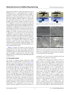
Figure 13 compares the behavior of the artificial hip
joint prosthesis between the experimental and simulated Figure 14. Scanning electron microscopy images of the fracture surface.
compression tests. The figure shows similar behavior of (A) Pure resin at ×250 magnification with 100 µm scale bar. (B) 1%
the artificial hip joint prosthesis between the experimental weight TiO at ×1,500 magnification with 10 µm scale bar. (C) 3% weight
2
TiO at ×1,500 × magnification with 10 µm scale bar. (D) 5% weight TiO
and the simulated compression test, with only a slight at ×1,500 magnification with 10 µm scale bar 2
2
difference in the fracture shape when the artificial hip joint
prosthesis breaks. nanoparticles and the resin, which is crucial for enhancing
3.3. Micrograph analysis the mechanical strength of the composite.
The results of scanning electron microscopy (SEM) Individual TiO nanoparticles should be visible as
2
indicate that the fracture surface of the specimen is smooth distinct entities in the SEM images, indicating proper
and neat in pure photopolymer as shown in Figure 14A. dispersion with minimal agglomeration. The presence of
After the addition of nanoparticles, the specimen surface individual nanoparticles contributes to strengthening the
looks rougher. In Figure 14B-D, there are clumps indicated composite and enhancing various functional properties.
by arrows; the larger the TiO nanoparticles used, the Although small aggregates of TiO nanoparticles form
2
2
more clumping and voids appear. Roughness on the when nanoparticles come into proximity and may be
fracture surface indicates an increase in the mechanical present in the SEM images, these aggregates do not form
strength of the specimen. When combining SLA resin large clusters or lumps. These aggregates offer improved
with TiO nanoparticles, several characteristic features are mechanical and functional properties comparable to
2
expected from the SEM images. SEM imaging provides individual nanoparticles. Besides, an adequate surface
detailed surface information, revealing the distribution, coverage of TiO nanoparticles on the resin matrix should
2
morphology, and interaction of nanoparticles within the also be observed in the SEM images. The nanoparticles
resin matrix. A uniform dispersion of TiO nanoparticles should be evenly distributed across the resin’s surface,
2
throughout the resin matrix should be observed in the SEM creating a continuous or semi-continuous layer. This
images, indicating a uniform dispersion and successful surface coverage ensures effective reinforcement and
incorporation of the nanoparticles into the resin. This desired functional properties, such as increased strength,
uniform dispersion signifies good interaction between the improved thermal stability, or enhanced optical properties.
Volume 4 Issue 3 (2025) 7 doi: 10.36922/MSAM025200032