
Page 89 - OR-1-2
P. 89
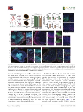
Figure 2. Derivation and characterization of regional FeBOs from different brain regions. The brightfield and immunofluorescence images of regional
            forebrain-derived  FeBOs  illustrate  the  expression  of  PAX6  (dorsal  forebrain  marker)  and  DLX2  (ventral  forebrain  marker).  Quantification  of  the
            efficiency of FeBO line establishment from dorsal and ventral forebrain regions is presented, and differences in mRNA expression of specific regional
            markers across varying culture ages are highlighted. In addition, the figure also demonstrates the reproducibility of marker expression in dorsal and
            ventral forebrain FeBOs across different lines.  Copyright © 2023 The author(s).
                                         19
            of cells to create 3D organoid models that closely resemble   Traditional methods of fetal stem cell isolation
            fetal tissues. These organoids can be cultured to represent   and  organoid  culture  often  depend  on  fetal  tissue
            various fetal organs, such as the brain, lungs, and kidneys,   samples obtained after the termination of pregnancy.
            providing a more accurate and physiologically relevant   This limitation restricts the investigation of fetal
            model of fetal development. By studying these organoids,   development and congenital diseases prenatally.
            researchers can gain insights into how genetic mutations   Therefore, there is a need for new patient-specific  in
            and environmental factors impact fetal tissues at the   vitro models to overcome these limitations.  AF, which
                                                                                                  20
            cellular and molecular levels. This method allows for early   contains cells from multiple developing fetal organs,
            and precise detection of genetic disorders such as Down   and tracheal fluid, which is closely related to fetal lung
            syndrome, cystic fibrosis, or congenital heart defects, all   development, offer alternative sources for such models.
            without the need for invasive procedures. In addition,   Through  single-cell  analysis technology, Gerli  et al.
                                                                                                            21
            AF-based organoids offer the potential for drug screening,   identified fetal epithelial stem/progenitor cells in both
            enabling the testing of therapeutic interventions in   amniotic and tracheal fluids and cultured tissue-specific
            models that closely mimic fetal development. Overall, this   organoids from these cells. Through RNA sequencing
            innovative technique represents a significant advancement   and immunostaining, the research team confirmed
            in prenatal diagnostics, providing a safer and more   the tissue specificity of the organoids (Figure  3). For
            comprehensive means of assessing fetal health.    example,  small  intestine  organoids  (SiAFOs)  exhibited


            Volume 1 Issue 2 (2025)                         4                            doi: 10.36922/OR025040005