
Page 39 - TD-2-1
P. 39
Tumor Discovery Breast cancer optical differentiation
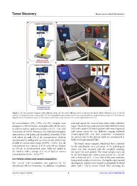
A B
C D
Figure 1. (A) The schematic diagram of the reflection setup. (B) The actual reflection setup to measure the tissue’s diffuse reflectance (R ): (1) the HS
d
camera, (2) the polychromatic source light, (3) the investigated breast samples, and (4) the computer and the image software analysis. (C) The schematic
diagram of the transmission setup. (D) The actual transmission setup to measure the tissue’s light transmission (T).
r
fat concentrations (3%, 1.75%, and 0%). Samples were read and signed the consent form before data collection
arranged in a 300 mL beaker, mixing the milk with the water began. The study was conducted from May 2020 to October
to achieve various lipid concentrations (0.1% ~ 1%, with 2021 with a total of 30 female patients who were diagnosed
increments of 0.05%). Moreover, the exploited absorption with breast cancer by two different imaging methods
material was a red Indian ink (Speedball, Statesville, USA) (mammogram/US) and had underwent mastectomy;
with whole fat milk (3% of fat concentration). Mixture the patient data for the present study are summarized in
was prepared by adding 200 mL from the ink with 0.2 mL Table S2 (Supplementary File).
of milk at concentration range (0.005% ~ 0.2%). The ink The breast tumor samples, which had been classified
concentration was constant at 0.1% with 200 µL dilution by the pathologists, were processed at the pathological
in 100 mL of demineralized water. Milk was added to center and then attached with the complete histology
the mixture with a syringe of 5 mL of lipid at various lab report for each patient. The breast samples were cut
concentrations (0.1% ~ 0.5%) [48,49] . by the pathologist into various sizes around 10 ~ 12 cm
2
with a thickness of 4 ~ 5 mm. One of the team members
2.4. Patient criteria and sample preparation
transported samples in an icebox. During the experimental
The current trial examination was approved by the investigation, samples were dipped in phosphate-buffered
Institutional Ethical Committee. In addition, all patients saline (pH = 7.4) to remove blood. After scanning, the
Volume 2 Issue 1 (2023) 4 https://doi.org/10.36922/td.258