
Page 48 - AIH-2-4
P. 48
Artificial Intelligence in Health ViT for neurodegeneration diagnosis
A B
C D
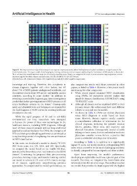
Figure 9. The mean attention maps of brain areas in our regions’ importance study, where the brightness of a pixel translates to its significance in the
model’s predictions. This figure results from merging the training, validation, and test datasets and feeding the final dataset of 580 samples to the model.
Then, we saved the model’s attention maps for all correctly classified scans. Finally, we computed the mean of saved attention maps to generate a mean
attention map for the whole dataset and each class. (A) CN, (B) MCI, (C) AD, (D) Overall.
Abbreviations: AD: Alzheimer’s disease; CN: Cognitively normal; MCI: Mild cognitive impairment.
knowledge and training. Therefore, this complexity in also compare our results with those presented in other
disease diagnosis, together with other factors, has left papers, as listed in Table 4. However, a few points worth
about 75% of NDD patients undiagnosed worldwide, and mentioning for a fair comparison:
this number rises to about 90% in low- and middle-income • While several papers examined NDD classification
countries, according to some studies. In addition to using DNNs, we exclusively selected studies that
3
revealing a considerable diagnosis gap, these investigations aimed for ternary classification (CN/MCI/AD) using
predict that the fast-growing number of NDD patients could 18 F-FDG PET scans
strain healthcare systems in the future. Consequently, • Although all chosen studies employed ADNI as their
3
novel and affordable tools and techniques are required for primary dataset, the authors may have used different
the final diagnosis of NDD and/or for assisting healthcare subsets to train and test the models
providers in this task. • As shown in Table 4, DNNs can surpass physicians
While the rapid progress of AI and its sub-fields when NDD diagnosis is solely based on brain
revolutionized our lives, researchers have attempted scans. However, domain experts usually consider
to harness the power of these new technologies in the a comprehensive collection of information for the
healthcare domain, including NDD diagnosis. Although clinical diagnosis, including the patient’s medical
most of these research projects utilized long-established history, genetics, blood tests, and cognitive and
approaches and architectures like CNNs, the emergence of physical evaluations. Consequently, instead of solely
ViTs and their groundbreaking performance convinced us relying on brain scans, doctors and nuclear medicine
to explore the potential of employing this new architecture physicians consider various factors, a practice that
in NDD classification. poses a substantial advantage over AI models.
In this work, we developed a model to classify F-FDG According to Table 4, in addition to a significantly
18
PET brain scans into CN, MCI, and AD. Specifically, higher F1 score, our model excels in distinguishing MCI
we designed the model based on the vanilla ViT-Base, cases, which proved to be the most challenging condition
introduced by Dosovitskiy et al., and trained on the ADNI to classify, compared to human experts and other models.
6
dataset. Combining the proposed data, pre-processing Apart from improving the model’s performance,
7
procedure, training recipe, and transfer learning enabled developing an explainable model was a pivotal goal of this
our model to achieve an F1 score of 81% (macro-average research. Therefore, we integrated the AAL3 brain atlas
of all classes), significantly outperforming previous information into the attention maps. This method resulted
approaches. To comprehend the model’s performance, we in a model that provides the brain region with the highest
Volume 2 Issue 4 (2025) 42 doi: 10.36922/AIH025140026