
Page 98 - AJWEP-22-6
P. 98
Li, et al.
Bi O at the (012) plane (JCPDF No.: 41-1449), ZrO The XRD patterns of the Ru-modified zeolites
3
2
2
at the (111) plane (JCPDF No.: 37-1484), and RuO at with varying initial molar ratios of Ru to ammonium
2
the (101) and (110) planes (JCPDF No.: 43-1027) were ion (Ru/NH₄⁺) are shown in Figure S2. All samples
observed. Interestingly, SbY-1 did not show detectable exhibited characteristic peaks of Y-zeolite along with
diffraction peaks corresponding to Sb oxide. However, the RuO₂ phase. As the Ru/NH₄⁺ ratio decreased, the
energy-dispersive X-ray spectroscopy (EDX) analysis intensity of the RuO₂ diffraction peaks also diminished,
(Figure S1B) confirmed the presence of Sb elements, indicating reduced loading of bulk RuO₂ with lower
suggesting a high degree of homogeneity and dispersion initial Ru content.
of Sb within the Y-zeolite matrix.
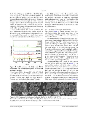
These results indicate that, except for SbY-1, the 3.1.2. SEM and TEM images of adsorbents
MY-1 adsorbents consist of two distinct phases: a The SEM (Figure 2) image exhibited that MY-1
MY-zeolite phase and a bulk metal oxide phase (MₓOᵧ). adsorbents retained the particle morphology of the
In contrast, SbY-1 appears to be a single-phase system original NH Y, exhibiting particles with crystal habits
4
with Sb ions uniformly dispersed within the zeolite. smaller than 0.9 µm.
The morphology and elemental distribution of MY-1
were further examined using SEM and EDX. The SEM
image exhibited that MY-1 adsorbents retained the
particle morphology of the original NH Y, exhibiting
4
particles with crystal habits smaller than 0.9 µm.
The SEM images of RuY-1 and ZrY-1 showed that
Y-zeolite particles were covered by bulk RuO and ZrO
2
2
nanoparticles, respectively. In contrast, the BiY-1 sample
exhibited particle conglutination. The EDX (Figure S1)
spectra confirmed the presence of Ru, Sb, Bi, and Zr
in their respective MY-1 samples. The metal content of
RuY-1, SbY-1, ZrY-1 and BiY-1 was 5.02, 13.85, 10.28
and 16.22 wt%, respectively (Table S1).
Low magnification TEM images of NH Y-zeolite
4
and the RuY-X series are shown in Figure 3. The
nano-rod-like particles of RuO with a width of
2
Figure 1. XRD patterns of NH Y and MY-1 30–40 nm and a length of 30–100 nm can be observed at
4
adsorbents. The characteristic peaks of the metal the surfaces of RuY-1 and RuY-1/2 samples. In contrast,
oxides are marked. RuO was not observed in RuY-1/4, RuY-1/8, and
2
Abbreviations: Bi₂O₃: Bismuth(III) oxide; MY: Metal RuY-1/16 samples, indicating a significant reduction in
ion-modified Y-zeolite; NH₄Y: Ammonium-form RuO content with decreasing initial Ru (Ru/NH mole
+
4
2
Y-zeolite; RuO₂: Ruthenium(IV) oxide; SbY: Antimony- ratio from 1 to 1/16). These observations are consistent
modified Y-zeolite; XRD: X-ray diffraction; Y-zeolite: with the XRD results (Figure S1). The high-resolution
Faujasite-type zeolite; ZrO₂: Zirconium(IV) oxide. TEM image of RuO is shown in the inset of Figure 3B,
2
A B C D
Figure 2. SEM images of adsorbents. (A) RuY-1. (B) SbY-1. (C) BiY-1. (D) ZrY-1.
Abbreviations: BiY: Bismuth-modified Y-zeolite; RuY: Ruthenium-modified Y-zeolite; SbY: Antimony-modified
Y-zeolite; SEM: Scanning electron microscopy; ZrY: Zirconium-modified Y-zeolite.
Volume 22 Issue 6 (2025) 92 doi: 10.36922/AJWEP025250204