Page 99 - AJWEP-22-6
P. 99

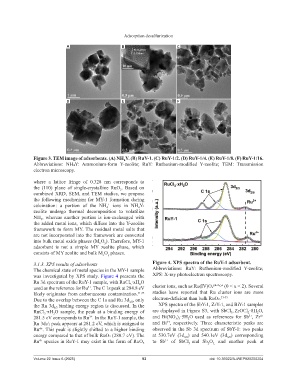
Adsorption desulfurization

                              A                        B                       C


                              D                        E                       F


                Figure 3. TEM image of adsorbents. (A) NH Y. (B) RuY-1. (C) RuY-1/2. (D) RuY-1/4. (E) RuY-1/8. (F) RuY-1/16.
                                                        4
                Abbreviations:  NH₄Y:  Ammonium-form  Y-zeolite;  RuY:  Ruthenium-modified  Y-zeolite;  TEM:  Transmission
                electron microscopy.

                where  a  lattice  fringe  of  0.328  nm  corresponds  to
                the  (110)  plane  of  single-crystalline  RuO .  Based  on
                                                      2
                combined XRD, SEM, and TEM studies, we propose
                the  following  mechanism  for  MY-1  formation  during
                calcination:  a portion of the NH  ions in NH Y-
                                                 +
                                                4
                                                              4
                zeolite  undergo thermal  decomposition  to volatilize
                NH ,  whereas  another  portion  is  ion-exchanged  with
                   3
                the added metal ions, which diffuse into the Y-zeolite
                framework to form MY. The residual metal salts that
                are not incorporated into the framework are converted
                into bulk metal oxide phases (M O ). Therefore, MY-1
                                             x
                                               y
                adsorbent  is  not  a  simple  MY  zeolite  phase,  which
                consists of MY zeolite and bulk M O  phases.
                                              x
                                                 y
                3.1.3. XPS results of adsorbents                    Figure 4. XPS spectra of the RuY-1 adsorbent.
                The chemical state of metal species in the MY-1 sample   Abbreviations:  RuY:  Ruthenium-modified  Y-zeolite;
                was investigated by XPS study. Figure 4 presents the   XPS: X-ray photoelectron spectroscopy.
                Ru 3d spectrum of the RuY-1 sample, with RuCl ·xH O
                                                              2
                                                          3
                used as the reference for Ru . The C 1s peak at 284.8 eV   cluster ions, such as Ru(IV)Oₓ⁽⁴⁻²ˣ⁾⁺ (0 < x < 2). Several
                                        3+
                likely originates from carbonaceous contamination. 31,32    studies have reported that Ru cluster ions are more
                Due to the overlap between the C 1s and Ru 3d , only   electron-deficient than bulk RuO₂. 33-35
                                                          3/2
                the Ru 3d binding energy region is discussed. In the   XPS spectra of the SbY-1, ZrY-1, and BiY-1 samples
                         5/2
                RuCl ·xH O  sample,  the  peak  at  a  binding  energy  of   are displayed in Figure S3, with SbCl , ZrOCl ·8H O,
                                                                                                       3
                                                                                                              2
                                                                                                                  2
                    3
                         2
                                                                                                               3+
                                                                                                                    4+
                281.5 eV corresponds to Ru . In the RuY-1 sample, the   and  Bi(NO ) ·5H O  used  as  references  for  Sb ,  Zr
                                        3+
                                                                              3 3
                                                                                    2
                                                                          3+
                Ru 3d₅/₂ peak appears at 281.2 eV, which is assigned to   and  Bi ,  respectively.  Three  characteristic  peaks  are
                Ru ⁺. This peak is slightly shifted to a higher binding   observed in the Sb 3d spectrum of SbY-1: two peaks
                  4
                energy compared to that of bulk RuO₂ (280.7 eV). The   at  530.7eV  (3d )  and  540.1eV  (3d )  corresponding
                                                                                  5/2
                                                                                                     3/2
                Ru ⁺ species in RuY-1 may exist in the form of RuOₓ   to Sb  of SbCl and Sb O   and  another  peak  at
                                                                          3+
                  4
                                                                                    3
                                                                                             2
                                                                                               3,
                Volume 22 Issue 6 (2025)                        93                           doi: 10.36922/AJWEP025250204
   94   95   96   97   98   99   100   101   102   103   104