
Page 20 - AN-1-3
P. 20
Advanced Neurology Early inhibition of PDK1 prevents AD-like pathology
numbers between Pdk1 cKO/5×FAD mice and their tdTomato+/NeuN+ cells in the cortex (Figure 1B). The
5×FAD littermates. P < 0.05 indicated a significant effect. cell counting results showed that ~92% of NeuN+ cells
were positive for tdTomato. Subsequently, we performed
3. Results fluorescence IHC for PDK1. We found that PDK1
3.1. Molecular analysis of the 5×FAD model with immunoreactivity was qualitatively reduced in the Pdk1
PDK1 deficiency through Emx1-Cre-mediated gene cKO/5×FAD cortex at 2.5 months compared with 5×FAD
recombination littermates (Figure 1C). Western blotting was performed for
PDK1 using cortical lysates from mice at 2.5 months. PDK1
It has been shown that long-term treatment of adult levels were notably decreased in the Pdk1 cKO/5×FAD
Tg2576 mice with BX912 decreases amyloid plaques but cortex compared with 5×FAD mice (Figure 1D), suggesting
causes lethal effect in mice . To investigate whether the efficient inactivation of PDK1 in Pdk1 cKO/5×FAD mice.
[22]
inactivation of PDK1, beginning from the embryonic stage,
could prevent AD-like pathology in 5×FAD mice , we 3.2. Plaque deposition is prevented in 5×FAD mice
[30]
generated Pdk1 ;Emx1-Cre/5×FAD (Pdk1 cKO/5×FAD) with PDK1 deficiency
f/f
mice, in which the deletion of PDK1 begins at embryonic To investigate whether PDK1 deletion affects plaque
day 10.5 (E10.5) in the forebrain neurons. The four different deposition in 5×FAD mice, IHC was performed for Aβ
genotypes are shown in Figure 1A. using brain sections from control, Pdk1 cKO, 5×FAD, and
To determine the expression pattern of Cre, Rosa26-LSL- Pdk1 cKO/5×FAD mice at three different ages (2.5, 4, and
tdTomato mice [16,47] were crossed to Emx1-Cre to generate 6 months). First, there were a few amyloid plaques in the
Emx1-Cre;Rosa26-LSL-tdTomato, in which tdTomato is 5×FAD cortex at 2.5 months but abundant amyloid plaques
expressed in Cre-positive (Cre+) cells. The brain sections at 4 and 6 months compared with non-Tg littermate controls
of the double mutant mice at P0 were used to perform (Figure 2A). In contrast, amyloid plaques were hardly seen
costaining for tdTomato and NeuN. We observed abundant in the Pdk1 cKO/5×FAD cortex at the aforementioned ages
A
B C
D
f/+
f/f
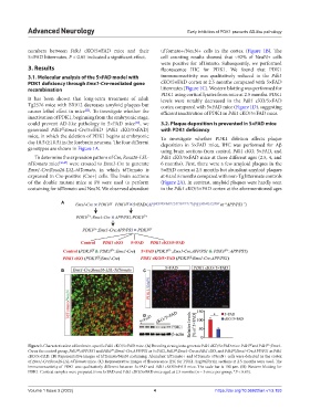
Figure 1. Characterization of forebrain-specific Pdk1 cKO/5×FAD mice. (A) Breeding strategies to generate Pdk1 cKO/5×FAD mice. Pdk1 and Pdk1 ;Emx1-
f/f
f/f
Cre as the control group, Pdk1 ;APP/PS1 and Pdk1 ;Emx1-Cre;APP/PS1 as 5×FAD, Pdk1 ;Emx1-Cre as Pdk1 cKO, and Pdk1 ;Emx1-Cre;APP/PS1 as Pdk1
f/f
f/+
cKO/5×FAD. (B) Representative images of tdTomato/NeuN costaining. Abundant tdTomato+ and tdTomato+/NeuN+ cells were detected in the cortex
of Emx1-Cre;Rosa26-LSL-tdTomato mice. (C) Representative images of fluorescence IHC for PDK1. Sagittal brain sections at 2.5 months were used. The
immunoreactivity of PDK1 was qualitatively different between 5×FAD and Pdk1 cKO/5×FAD mice. The scale bar is 100 μm. (D) Western blotting for
PDK1. Cortical samples were prepared from 5×FAD and Pdk1 cKO/5×FAD mice aged at 2.5 months (n = 3 mice per group; *P < 0.05).
Volume 1 Issue 3 (2022) 4 https://doi.org/10.36922/an.v1i3.153