
Page 22 - AN-1-3
P. 22
Advanced Neurology Early inhibition of PDK1 prevents AD-like pathology
A
B C
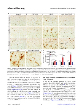
Figure 3. Diminished microgliosis in Pdk1 cKO/5×FAD mice. (A) Representative images of IHC for Iba1 in the cortex. Brain sections were prepared from
control, Pdk1 cKO, 5×FAD, and Pdk1 cKO/5×FAD mice aged 2.5, 4, and 6 months. The immunoreactivity of Iba1 was qualitatively different between the
control and 5×FAD mice. The scale bar is 20 μm. (B) Enlarged images for boxed regions in (A). The scale bar is 25 μm. (C) Quantification results showing
the average number of Iba1+ cells in the cortex. There were significant differences in the number of Iba1+ cells between 5×FAD and Pdk1 cKO/5×FAD
mice at 2.5 (*P < 0.05), 4 (***P < 0.001), and 6 (***P < 0.001) months (n = 3–4 mice per group per age).
To study whether there are changes to astrocytes in 3.4. mTOR signaling is inhibited in 5×FAD mice with
Pdk1 cKO/5×FAD mice, IHC was performed for GFAP. PDK1 deficiency
First, it was observed that GFAP immunoreactivity was In the mTOR signaling pathway, S6 kinase (S6K)
qualitatively higher in the Pdk1 cKO cortex and the 5×FAD phosphorylates S6 at Ser235 and Ser236 (Ser235/236)
cortex at 2.5, 4, and 6 months compared with the littermate following its activation by mTOR [49,50] . We investigated
controls (Figure 4A). GFAP immunoreactivity was reduced whether this pathway is affected by PDK1 deletion. First,
in the Pdk1 cKO/5×FAD cortex compared with the 5×FAD we performed Western blotting for pS6 Ser235/236 . We found
cortex (Figure 4A and B). Second, the total number of that the levels of pS6 Ser235/236 were significantly lower in
GFAP+ cells in the 5×FAD cortex at 4 or 6 months was the Pdk1 cKO/5×FAD cortex than in the 5×FAD cortex
more than that in the control cortex (Figure 4C; P < 0.001), (Figure 5A and B). Second, we performed Western blotting
but it was smaller in the Pdk1 cKO/5×FAD cortex than in for phosphorylated 4E-BP1, an important member of the
the 5×FAD cortex (Figure 4C; P < 0.001). These results mTOR pathway . We found that the levels of p4E-BP1 Ser65
[51]
suggest that astrogliosis may be attenuated by PDK1 were lower in the Pdk1 cKO/5×FAD cortex compared with
deletion in 5×FAD mice. 5×FAD mice (Figure 5A and B). Third, we performed IHC
Volume 1 Issue 3 (2022) 6 https://doi.org/10.36922/an.v1i3.153