
Page 87 - AN-2-2
P. 87
Advanced Neurology Brain AT -R and kidney crosstalk
1
stainless steel screw was anchored to the skull. Cannulae or artificial cerebral fluid (ACF), inserting a stainless steel
were placed 2 mm above the final place of injection. injection cannula (30 gauge) into the guide cannula. The
According to Paxinos and Watson Atlas , coordinates for cannula was attached through a polyethylene catheter
[26]
lateral ventricle concerning bregma were: Anteroposterior (P20) to a 25 µL microsyringe (Hamilton). Volumes of
(AP) = –0.92 mm, L = ±1.5 mm, dorsoventral 1 µL of ACF or Los solution were gradually injected over a
(DV) = –6.8 mm. The cannulae were implanted in the right 1-min period into the left and right sides using an infusion
and left sides of the lateral ventricle. bomb (HARVARD, model 22). The injection cannula was
In the anesthetized animals, the surgical denervation left in place for an additional 20 s to allow complete liquid
was made under a magnifier. First, the right kidney was diffusion [23,31,32] .
exteriorized, the lipid and connective tissue from vessels 2.6. Biochemistry
at the renal pelvis level was separated, and the nerve was
dissected, leaving the renal artery and vein intact. The 2.6.1. Urine collection and blood samples
denervation phenol 10% (neurolytic) was applied to make The daily urine production was collected and registered,
it more accurate. The kidney was introduced, and the same and 12 h of urine production was collected after Los/ACF
procedure was performed in the left kidney. Finally, the injection and centrifuged at 10,000 G for 10 min. The
surgery was finished with muscle and skin sutures [27-30] . The supernatant was kept at –20°C until analysis.
sham animals underwent the same procedure until renal
nerve visualization for each kidney. The renal denervation At the end of the experiment, blood samples were taken
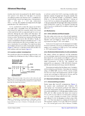
was verified in kidney slices with a hematoxylin-eosin stain from the heart’s left ventricle in anesthetized animals. The
(Figure 2). Moreover, renal denervation did not affect the samples were centrifuged at 3000 rpm for 5 min and kept
o
food, sodium, and water intake (Figure 3). at –80 C until analysis (Figure 1).
2.5. Losartan cerebral microinjection 2.6.2. Biochemical determinations in urine and plasma
The animals were placed in metabolic cages from day 1 and The urinary volume was registered, and the parameters
were bilaterally administered on day 5 with Los (4 µg/1 µL) such as creatinine, osmolarity, sodium, and potassium were
determined. The urine samples were diluted in water 1:20
and analyzed for creatinine using a modified Jaffe reaction
under spectrometry at 510 nm. The osmolarity was
measured by an osmometer 3.300 ADVANCE (System),
and the ions were measured using a selective electrode.
The parameters analyzed in the plasma were: Creatinine,
sodium potassium, and chlorine. Creatinine was measured
using a modified Jaffe reaction. Briefly, plasma samples
(0.5 mL) were mixed with trichloroacetic acid 5% in equal
volume and centrifuged at 5000 rpm for 10 min, and the
ions were measured using a selective electrode.
Figure 1. Experimental design: 6-day protocol with normosodic and
hypersodic diet. The stereotaxic surgery was performed at the beginning 2.7. Neuronal activation analysis
of the protocol, and vehicle/losartan was administered on day five. Twelve
hours later, the animals were sacrificed. 2.7.1. Immunostaining procedure
Twelve hours after Los/ACF administration (Figure 1),
A B the animals were anesthetized with Urethane 50%
(100 mg/kg) and transcardially perfused with 200ml of
0.9% saline and heparin (200 µL/L), followed by 200ml of
4% paraformaldehyde in 0.1M phosphate-buffered saline
(PBS, pH 7.4). After removal, the brains were stored at
4°C in a 30% sucrose solution. Coronal sections of 40 µm
were cut using a freezing microtome (Leica CM1510S)
and collected in 0.01 M phosphate-buffered saline (PBS,
Figure 2. Representative microphotographs of histological sections
of the kidney. (A) SHAM, renal nerve intact, (B) RDN, renal nerve pH = 7.4). They were placed in a mixture of 10% H O and
2
2
lesioned. Renal nerve lesion verification in hematoxylin-eosin stained 10% methanol, washed 3 times with PBS 0.01 M, and then
×100 sections. in a 10% normal goat serum (NGS; Natocor, Córdoba,
Volume 2 Issue 2 (2023) 3 https://doi.org/10.36922/an.393