
Page 88 - AN-2-2
P. 88
Advanced Neurology Brain AT -R and kidney crosstalk
1
A
B
C
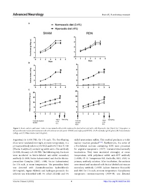
Figure 3. Food, sodium, and water intake in rats treated with a 0.4% normosodic diet (white) and with a 4% hipersodic diet (black) for 5 days prior to
intracerebroventricular administration with vehicle/losartan (left panel: SHAM) and (right panel: RDN). (A) Food intake (g/100 g/bw), (B) Sodium intake
(mEq), and (C) Water intake (mL/100 g/bw).
Argentina) in 0.1M PBS, for 2 h each. The free-floating nickel ammonium sulfate. This method produces a violet
slices were incubated overnight, at room temperature, in a nuclear reaction product [33,34] . Furthermore, the series of
primary antibody solution (2% NGS and 0.3% Triton X-100 c-Fos-labeled sections containing SON were processed
(Flucka Analytical)) containing rabbit anti-c-Fos antibody for arginine-vasopressin (AVP) immunohistochemically
(1:3000; Abcam), in 0.1M PBS. The following day, the slices localization. They were incubated overnight, at room
were incubated in biotin-labeled anti-rabbit secondary temperature, with polyclonal rabbit anti-AVP antibody
antibody (1:1000; Vector Laboratories) and Avidin-Biotin- (1:3000; PS 41 Vasopressin-NP; Rockville, MD, USA) in
peroxidase Complex (ABC; 1:500; Vector Laboratories) primary antibody solution. After incubation, the sections
for 2 h each, at room temperature. The peroxidase label were rinsed and incubated with biotin-labeled anti-mouse
was detected with diaminobenzidine hydrochloride secondary antibody (1:3000 Jackson Immune Research)
(0.5 mg/mL, Sigma-Aldrich) and hydrogen peroxide; the and ABC for 2 h each, at room temperature. Cytoplasmic
solution was intensified with 1% cobalt chloride and 1% vasopressin immunoreactivity (AVP-IR) was detected
Volume 2 Issue 2 (2023) 4 https://doi.org/10.36922/an.393