
Page 104 - AN-3-3
P. 104
Advanced Neurology Proteomic analysis of microglia exosomes
A B C
D
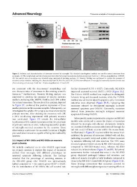
Figure 2. Isolation and characterization of exosomes secreted by microglia. The ultrafast centrifugation method was used to extract exosomes from
microglia. (A) The morphology and size of exosomes were observed using transmission electron microscopy. Scale bar = 100 nm; magnification: ×100000.
(B) The particle size of exosomes was detected using nanoparticle tracking analysis. (C) Western blotting was performed to analyze the presence of
exosome surface markers, including Alix, heat shock protein70, Flotillin, and CD9. (D) The internalization of microglial exosomes by neurons in vitro was
observed using a microscope. Scale bar = 50 μm; magnification: ×10 and ×40.
are consistent with the documented morphology and further decreased (0.335 ± 0.027). Conversely, M2-EXOs
size characteristics of exosomes in the existing scientific improved neuronal survival (0.652 ± 0.020 [Figure 3A]).
literature. Furthermore, Western blotting analysis was The Calcein AM/PI method was employed to distinguish
17
employed to confirm the presence of specific exosome between living and deceased neurons, while the TUNEL
markers, including Alix, HSP70, Flotillin, and CD9, within method was utilized to identify apoptotic cells. Consistent
the isolated exosomes. The results of this analysis, depicted outcomes were observed (Figure 3B-E), indicating that
in Figure 2C, confirmed the positive expression of these exosomes released by detrimental microglia increased
marker proteins in the exosome samples. Subsequently, we neuronal apoptosis post-OGD/R. Conversely, exosomes
investigated the uptake of microglia-secreted exosomes by derived from protective microglia mitigated neuronal
primary neurons. After staining the exosomes with Dil, apoptosis following OGD/R.
a 24-h coculturing experiment with primary neurons
was conducted. Figure 2D reveals the intracellular Subsequently, animal experiments using mouse tMCAO
localization of Dil-stained exosomes within the cytoplasm models were conducted to assess the impact of exosomes
of primary neurons in vitro, indicating successful uptake released by microglia with distinct phenotypes. Initially,
of microglia-secreted exosomes by the neurons. These we examined whether exosomes administered through the
observations underscore the successful isolation of highly tail vein could infiltrate neurons within the mouse brain.
pure and intact exosomes capable of being internalized by As illustrated in Figure 3F, neurons within the mouse brain
neurons. exhibited the presence of exosomes labeled with red dye,
indicating successful entry and uptake of these exosomes
3.3. Impact of M1-EXOs and M2-EXOs on neurons by neurons following tail vein injection. The TTC assay
post-ischemia revealed a greater infarct volume in M1-EXO-treated mice
We initially conducted an in vitro OGD/R experiment compared to M0-EXO-treated mice, whereas M2-EXO
on primary neurons to explore the impact of exosomes reduced the infarct volume (Figure 3G and H). Behavioral
derived from microglia with different phenotypes on assessments further indicated that mice in the M2-EXO
neuronal apoptosis following ischemia. The findings group displayed prolonged balance rod retention and
revealed that the percentage of surviving neurons in enhanced muscle strength in both forelimbs compared to
the M0-EXO group after OGD/R was approximately those in the M0-EXO-treated tMCAO group (Figure 3I-K).
halved compared to = the control group (0.497 ± 0.029). These findings substantiate that exosomes secreted by
On the introduction of M1-EXOs, the cell survival rate microglia with different phenotypes exert varying effects on
Volume 3 Issue 3 (2024) 6 doi: 10.36922/an.3166