
Page 62 - ARNM-2-3
P. 62
Advances in Radiotherapy
& Nuclear Medicine NCRT for T3N0M0 ESCC
+ S had an increased risk of death (HR = 10.99; 95% 3.5. Failure patterns in the whole population
CI = 1.00 – 121.25), while in other subgroups, NCRT At the final follow-up, the total number of failures
+ S reduced the risk of death (Table 3). Patients with (comprising local recurrences, distant metastases, and
tumors located in the lower segment (HR = 1.17; 95% deaths) was 228/443 (51.5%). The NCRT + S and S
CI = 0.65 – 2.12), tumor length >5 cm (HR = 1.54; groups included 36 and 192 patients, respectively. Of
95% CI = 0.37 – 6.39), or with vascular invasion had the 103 (103/443, 23.3%) local recurrences, 84 were
a significantly increased risk of progression when intrathoracic local-regional recurrences, 12 were in the
undergoing NCRT + S (Table 4).
supraclavicular LNs, and seven were in the intraabdominal
LNs. The NCRT + S group, with a rate of 14 (14/100), showed
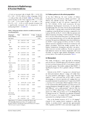
Table 3. Subgroup analysis related to overall survival in the a significant reduction in local recurrence compared to the
overall population
S group (89/343, 25.9%, P = 0.013). Fifty-two patients had
Subgroup S Total HR (95% CI) P‑value P‑value for distant metastases, including 13 with metastasis in the lung,
versus NCRT+S interaction four in the liver, three in the bone, three in the brain, 24 with
Age (years) 0.939 two or more metastatic sites, and five with other metastatic
≤65 269 0.58 (0.35 – 0.99) 0.045 locations. No significant differences were observed between
>65 174 0.59 (0.30 – 1.19) 0.142 the NCRT + S and S groups (11/100 vs. 41/343, P = 0.794).
Sex 0.174 Thirteen patients experienced both local recurrences and
distant metastases. Thirty-one deaths occurred due to
Male 360 0.60 (0.39 – 0.92) 0.020 fistulas, hematemesis, hemoptysis, and other non-tumor-
Female 83 0.19 (0.03 – 1.37) 0.098 related internal medical conditions. Twenty-one patients
KPS 0.181 had unknown reasons for disease progression and cause
≥90 376 0.64 (0.41 – 1.01) 0.055 of death; in contrast, eight patients developed a second
80 67 0.30 (0.09 – 0.98) 0.046 primary tumor. The results are presented in Table 5.
Weight loss 0.482
Yes 170 0.67 (0.32 – 1.40) 0.287 4. Discussion
No 273 0.51 (0.31 – 0.84) 0.009 This study introduced a novel approach to evaluating
Tumor location 0.563 patients with pre-T3N0M0 status and is the first to compare
Upper 120 0.51 (0.20 – 1.29) 0.154 survival outcomes between NCRT + S and surgery alone in
this specific group. The results confirmed that NCRT + S
Middle 218 0.46 (0.20 – 1.05) 0.065 significantly improved OS in patients with pre-T3N0M0
Lower 105 0.73 (0.38 – 1.40) 0.340 status.
Tumor length 0.74
(cm) Patients in the NCRT + S group were selected based
≤5 354 0.56 (0.36 – 0.87) 0.009 on their pretreatment clinical T stage (cT) rather than
the post-operative pathological T (pT) stage. Previous
>5 86 0.85 (0.12 – 6.21) 0.872 studies 4,5,11 have confirmed that NCRT leads to tumor
Unknown 3 NA (NA – NA) NA downstaging, so the post-operative pT staging does not
LVI 0.045 accurately represent the pretreatment T stage in the NCRT
Yes 24 10.99 (1.00 – 121.25) 0.050 + S group. Since endoscopic ultrasonography (EUS) and
No 419 0.54 (0.35 – 0.82) 0.004 CT are widely used in clinical practice, this study utilized cT
PNI 0.719 staging determined through EUS and CT. Both Lightdale
19
20
Yes 86 0.65 (0.25 – 1.65) 0.362 and Kulkarni and a meta-analysis reported that the
accuracy of EUS in diagnosing cT3 tumors in esophageal
No 357 0.56 (0.35 – 0.90) 0.016 cancer was ≥90%. The accuracy of CT in distinguishing
Total lymph 0.435 T3 lesions was 86.7%. Therefore, the selection of patients
21
nodes excised with cT3 tumors in this study’s NCRT + S group closely
<18 202 0.43 (0.25 – 0.73) 0.002 represented the actual pretreatment T3 status.
≥18 241 0.68 (0.35 – 1.32) 0.257 21
Due to the limited accuracy of CT in LN staging,
Abbreviations: CI: Confidence intervals; HR: Hazard ratio; clinical N (cN) staging before treatment is often less
KPS: Karnofsky Performance Status; LVI: Lymphovascular
12
invasion; NCRT+S: Neoadjuvant chemoradiotherapy plus surgery; reliable. A previous study reported that approximately
PNI: Perineural invasion; S: Surgery. 50% of patients with ESCC who underwent surgery alone
Volume 2 Issue 3 (2024) 7 doi: 10.36922/arnm.3821