
Page 79 - ARNM-2-3
P. 79
Advances in Radiotherapy
& Nuclear Medicine OrthoCT experimental proof of concept
include (1) formation of edema in the irradiated area,
(2) tumor regression/progression, (3) filling of cavities
with edematous tissue (e.g., due to inflammation),
(4) change in tissue permeability, (5) weight loss/gain, and
(6) misalignment of patient positioning, among others. 2-6
Image-guided radiation therapy (IGRT) allows for
more precise tumor targeting, thereby reducing the side
effects of eventual morphological and/or anatomical
changes. Cone-beam computed tomography is one of
the most commonly used IGRT techniques for treatment
monitoring, as it provides visualization of the target with
volumetric imaging and relatively high-resolution soft-
7
tissue information. However, this technique results in
10
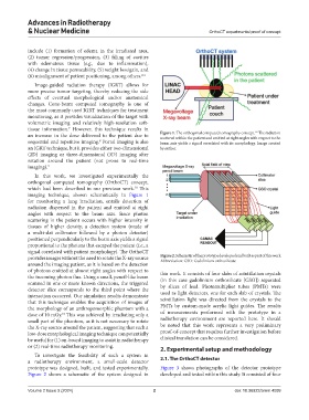
an increase in the dose delivered to the patient due to Figure 1. The orthogonal computed tomography concept. The radiation
scattered within the patient and emitted at right angles with respect to the
sequential and repetitive imaging. Portal imaging is also beam axis yields a signal correlated with its morphology. Image created
8
an IGRT technique, but it provides either two-dimensional by author.
(2D) imaging or three-dimensional (3D) imaging after
rotation around the patient (not prone to real-time
imaging). 9
In this work, we investigated experimentally the
orthogonal computed tomography (OrthoCT) concept,
which had been described in our previous work. This
10
imaging technique, shown schematically in Figure 1
for monitoring a lung irradiation, entails detection of
radiation dispersed in the patient and emitted at right
angles with respect to the beam axis. Since photon
scattering in the patient occurs with higher intensity in
tissues of higher density, a detection system (made of
a multi-slat collimator followed by a photon detector)
positioned perpendicularly to the beam axis yields a signal
proportional to the photons that escaped the patient (i.e., a
signal correlated with patient morphology). The OrthoCT
provides images without the need to rotate the X-ray source Figure 2. Schematic of the prototype developed and built as part of this work
Abbreviation: GSO: Gadolinium orthosilicate.
around the imaging patient, as it is based on the detection
of photons emitted at almost right angles with respect to this work. It consists of four slabs of scintillation crystals
the incoming photon flux. Using a small, pencil-like beam (in this case gadolinium orthosilicate [GSO]) separated
scanned in one or more known directions, the triggered by slices of lead. Photomultiplier tubes (PMTs) were
detector slice corresponds to the third point where the used as light detectors, one for each slab of crystals. The
interaction occurred. Our simulation results demonstrate
that this technique enables the acquisition of images of scintillation light was directed from the crystals to the
the morphology of an anthropomorphic phantom with a PMTs by custom-made acrylic light guides. The results
dose of 10 mGy. This was achieved by irradiating only a of measurements performed with the prototype in a
10
small part of the phantom, as it is not necessary to rotate radiotherapy environment are reported here. It should
the X-ray source around the patient, suggesting that such a be noted that this work represents a very preliminary
low-dose morphological imaging technique can potentially proof-of-concept that requires further investigation before
be useful for (1) on-board imaging to assist in radiotherapy clinical translation can be considered.
or (2) real-time radiotherapy monitoring. 2. Experimental setup and methodology
To investigate the feasibility of such a system in
a radiotherapy environment, a small-scale detector 2.1. The OrthoCT detector
prototype was designed, built, and tested experimentally. Figure 3 shows photographs of the detector prototype
Figure 2 shows a schematic of the system designed in developed and tested within this study. It consisted of four
Volume 2 Issue 3 (2024) 2 doi: 10.36922/arnm.4099