
Page 84 - ARNM-2-3
P. 84
Advances in Radiotherapy
& Nuclear Medicine OrthoCT experimental proof of concept
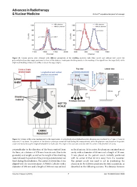
Figure 10. Counts spectra were obtained with different geometries of the shielding material, with (blue curve) and without (red curve) the
polymethylmethacrylate target positioned in front of the detector. Inadequate shielding results in the masking of the signal from the target (left), while
improved shielding enhances the ability to detect the target (right).
Figure 11. Scheme of the setup implemented in the experiments. A cylindrical polymethylmethacrylate phantom was irradiated by a 5 mm × 5 mm (at
isocenter) X-ray beam. The position of the detector prototype remained fixed during the measurements, while the phantom was placed on the patient
couch and moved to acquire longitudinal and/or vertical scans. The origin of the scan axis coincides with the center of the phantom air cavity.
perpendicular to the direction of the beam emitted from as the phantom. At its center, the phantom contained an air
the linac, at a distance of 170 mm from its axis. Due to its cavity with a diameter of 40 mm and a length of 20 mm.
geometry and weight, as well as the weight of the shielding It was placed on the patient couch, initially positioned
material used, the position of the prototype system remained with its center shifted 60 mm away from the isocenter.
fixed during the irradiation. The center of crystal line 1 was The patient couch was used to aid in positioning the
aligned with the isocenter plane. A PMMA cylinder with a phantom in the different positions for data acquisition, as
diameter of 180 mm and a length of 300 mm was utilized described in the following sections. The distance between
Volume 2 Issue 3 (2024) 7 doi: 10.36922/arnm.4099