
Page 222 - EJMO-9-3
P. 222
Eurasian Journal of
Medicine and Oncology FN3K–Nrf2 axis inhibition in breast cancer
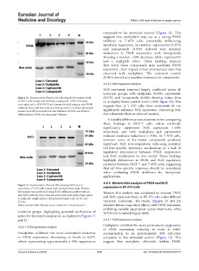
compared to the untreated control (Figure 11). This
suggests that oxaliplatin may act as a strong FN3K
inhibitor in T-47D cells, potentially influencing
metabolic regulation. In contrast, capivasertib (0.979)
and lansoprazole (0.876) induced only minimal
reductions in FN3K expression, with lansoprazole
showing a modest ~10% decrease, while capivasertib
had a negligible effect. These findings indicate
that while these compounds may modulate FN3K
expression, their impact is less pronounced than that
observed with oxaliplatin. The untreated control
(0.967) served as a baseline reference for comparison.
3.4.2.2. Nrf2 expression analysis
Nrf2 expression remained largely unaffected across all
treatment groups, with oxaliplatin (0.909), capivasertib
Figure 11. Representative Western blot showing FN3K protein levels (0.979), and lansoprazole (0.886) showing values close
in T47D cells treated with different compounds. T47D cell lysates to or slightly below control levels (1.049; Figure 12). This
were subjected to SDS-PAGE and immunoblotted using an anti-FN3K suggests that, in T-47D cells, these compounds do not
antibody. Beta actin was used as a loading control. A molecular weight significantly influence Nrf2 expression, contrasting with
marker (lane M) is included for size reference (29 kDa and 45 kDa)
Abbreviations: FN3K: Fructosamine-3-kinase their observed effects in other cell models.
A notable difference was observed when comparing
these findings to MCF-7 cells, where amiloride
significantly suppressed Nrf2 expression (~60%
reduction), and both oxaliplatin and capivasertib
induced moderate reductions (~20%). In T-47D cells,
however, none of the tested compounds produced
significant Nrf2 downregulation, indicating potential
cell-line-specific resistance mechanisms or a lack of
regulatory dependency between FN3K suppression
and Nrf2 modulation in this model. These findings
highlight differences in FN3K and Nrf2 regulatory
pathways between MCF-7 and T-47D cells, suggesting
that cell-line-specific responses should be considered
when evaluating FN3K inhibitors for therapeutic
applications.
3.4.3. Western blot analysis of FN3K and Nrf2
Figure 12. Representative Western blot showing Nrf2 protein
expression in T47D cells treated with selected compounds. Western expression in BT-474 Cells
blot analysis was performed using T47D cell lysates probed with an Western blot analysis was conducted to evaluate FN3K
anti-Nrf2 antibody. Beta actin was used as the internal loading control. and Nrf2 expression levels in BT-474 cells under different
A molecular weight marker (M) indicates bands at 29, 45, 67, and
97 kDa treatment conditions. The results (Figures 13 and 14)
Abbreviations: Nrf2: Nuclear factor erythroid 2-related factor 2 illustrate distinct regulatory effects, with FN3K expression
exhibiting variable suppression across treatments, while
treatment groups, highlighting potential mechanisms of Nrf2 levels remained largely stable.
action for the tested compounds, as depicted in Figures 11
and 12. 3.4.3.1. FN3K expression analysis
Oxaliplatin exhibited the most pronounced suppression
3.4.2.1. FN3K expression analysis
of FN3K expression, reducing its levels to 0.681,
Oxaliplatin exhibited the most substantial reduction corresponding to an approximately 30% reduction
in FN3K expression, decreasing its levels to 0.677, compared to the untreated control (Figure 13). This
which representing approximately a 30% suppression suggests that oxaliplatin effectively inhibits FN3K,
Volume 9 Issue 3 (2025) 214 doi: 10.36922/EJMO025150114