
Page 120 - GTM-4-3
P. 120
Global Translational Medicine Rapid diagnostic imaging on biopsy needle
A
B C
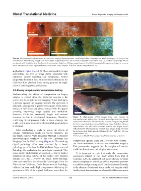
Figure 6. Microscopy with ultraviolet surface excitation imaging of a mouse tumor model obtained via a 14-gauge core needle biopsy gun. (A) Image of the
mouse tumor, stitched using ImageJ. Scale bar: 500 µm; magnification: 10×, (B) Zoomed-in segment of the tumor from (A), stitched using ImageJ. Nuclei
are stained with Hoechst with a Rhodamine B counterstain. Scale bar: 500 µm; magnification: 10×, (C) Color-adjusted image using ImageJ to improve
visual comparison between pig breast biopsies and mouse tumor biopsies. Scale bar: 500 µm; magnification: 10×.
applications (Figure 7A and B). These comparative images A
demonstrate the level of image clarity achievable with
optimized sample handling and preparation, further
supporting the notion that, with continued refinement, the
CoreView ION platform holds strong promise for rapid,
point-of-care diagnostic applications.
3.3. Biopsy integrity under compression testing
Understanding the effects of compression on biopsy B
samples is critical since the prototype requires a flat
surface for planar microscopic imaging. When the biopsy
is pressed against the imaging window, the specimen is
flattened, allowing for a greater percentage of the tissue
surface in full focus and direct contact with the quartz
coverslip, improving image quality and resolution.
However, CNBs are structurally fragile, and external
stressors can lead to mechanical breakdown. Therefore, Figure 7. High-quality MUSE images from core biopsies in
confirming if compression leads to tissue damage that non-needle-based applications. (A) Fresh normal prostate core biopsy
could compromise downstream histopathological analysis stained with alternative Rhodamine and Hoechst, imaged using MUSE
by the Levenson Lab, University of California, Davis. Scale bar: 500 µm;
is essential. magnification: 10×, (B) Fresh cancerous prostate core biopsy stained
After conducting a study to assess the effects of with alternative Rhodamine and Hoechst, also imaged using MUSE by
22
the Levenson Lab, University of California, Davis. Scale bar: 500 µm;
varying compression levels on biopsy integrity, the magnification: 10×.
pig breast samples were processed through a standard Abbreviation: MUSE: Microscopy with ultraviolet surface excitation.
histopathological workflow at the UW Histology and
Imaging Core for H&E staining (Figure 8). The resulting compression applied using the CoreView ION system—
digital pathology slides were reviewed by a breast the tissue maintained structural and molecular integrity.
pathology specialist from the UW Medicine Department of This preservation suggests that the samples will remain
Pathology. On evaluation, the pathologist remarked, “The viable for downstream pathological assessment.
tissue quality and staining are excellent… able to make a It is important to note that while the compression
diagnosis on tissue samples of this quality” (Dr. Suzanne mechanism utilized in this study differs from that of
Dintzis, MD, PhD, October 28, 2022). These findings, CoreView ION, the employed test fixture allowed for more
later confirmed by a board-certified pathologist from the precise compression control, as well as improved specimen
University of California, Davis, Department of Pathology, accessibility and processing speed. One limitation of this study
indicate that even under high compression—flattening the is the absence of cancerous tissue in the test samples; while
specimen to 30% of its original thickness, more than the healthy tissue demonstrated no observable differences across
Volume 4 Issue 3 (2025) 112 doi: 10.36922/GTM025170039