
Page 119 - GTM-4-3
P. 119
Global Translational Medicine Rapid diagnostic imaging on biopsy needle
3. Results and discussion Seattle, WA, were utilized as additional specimens. The
tumor images were captured using the CoreView prototype
3.1. MUSE imaging of porcine tissues after biopsy acquisition and staining (Figure 6).
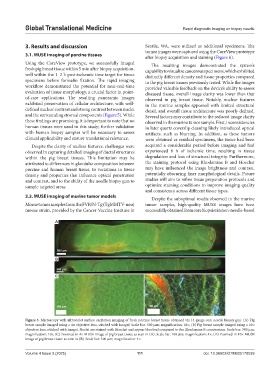
Using the CoreView prototype, we successfully imaged The resulting images demonstrated the system’s
fresh pig breast tissue within 5 min after biopsy acquisition, capability to visualize cancerous specimens, which exhibited
well within the 1–2 h post-ischemic time target for tissue distinctly different density and tissue properties compared
specimens before formalin fixation. The rapid imaging to the pig breast tissues previously tested. While the images
workflow demonstrated the potential for near-real-time provided valuable feedback on the device’s ability to assess
evaluation of tissue morphology, a crucial factor in point- diseased tissue, overall image clarity was lower than that
of-care applications. The resulting panoramic images observed in pig breast tissue. Notably, nuclear features
exhibited preservation of cellular architecture, with well- in the murine samples appeared with limited structural
defined nuclear contrast and strong contrast between nuclei detail, and overall tissue architecture was poorly defined.
and the surrounding stromal components (Figure 5). While Several factors may contribute to the reduced image clarity
these findings are promising, it is important to note that no observed in the murine tumor sample. First, inconsistencies
human tissues were used in this study; further validation in later quartz coverslip cleaning likely introduced optical
with human biopsy samples will be necessary to assess artifacts, such as blurring. In addition, as these tumors
clinical applicability and ensure translational relevance. were obtained as residual specimens, the tissue had been
Despite the clarity of nuclear features, challenges were acquired a considerable period before imaging and had
observed in capturing detailed imaging of ductal structures experienced 6 h of ischemic time, resulting in tissue
within the pig breast tissues. This limitation may be degradation and loss of structural integrity. Furthermore,
attributed to differences in glandular composition between the staining protocol using Rhodamine B and Hoechst
porcine and human breast tissue, to variations in tissue may have influenced the image brightness and contrast,
density and properties that influence optical penetration potentially obscuring finer morphological details. Future
and contrast, and to the ability of the needle biopsy gun to studies will aim to refine tissue preparation protocols and
sample targeted areas. optimize staining conditions to improve imaging quality
and consistency across different tissue types.
3.2. MUSE imaging of murine tumor models
Despite the suboptimal results observed in the murine
Mouse tumor samples from the FVB/N-Tg (TgMMTV-neu) tumor samples, high-quality MUSE images have been
mouse strain, provided by the Cancer Vaccine Institute in successfully obtained from core biopsies in non-needle-based
A
B
C
Figure 5. Microscopy with ultraviolet surface excitation imaging of fresh porcine breast tissue obtained via 14-gauge core needle biopsy gun. (A) Pig
breast sample imaged using a 4× objective lens, stitched with ImageJ. Scale bar: 500 µm; magnification: 10×, (B) Pig breast sample imaged using a 10×
objective lens, stitched with ImageJ. Nuclei are stained with Hoechst and appear blue/teal compared to the Rhodamine B counterstain. Scale bar: 500 µm;
magnification: 10x, (C) Zoomed-in 4× MUSE image of pig breast tissue as seen in (A). Scale bar: 100 µm; magnification: 4×, (D) Zoomed-in 10× MUSE
image of pig breast tissue as seen in (B). Scale bar: 100 µm; magnification: 4×.
Volume 4 Issue 3 (2025) 111 doi: 10.36922/GTM025170039