
Page 121 - GTM-4-3
P. 121
Global Translational Medicine Rapid diagnostic imaging on biopsy needle
compression levels, diseased tissue may respond differently. contrast and edge sharpness. In the MUSE image, nuclei
Further studies incorporating malignant samples are necessary appear brighter than the surrounding stroma, whereas
to evaluate potential compression-induced artifacts. in the H&E image, nuclei are darker. Intensity profiles
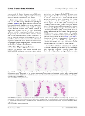
Murine tissue sections were also submitted to the across representative nuclei demonstrate this inverse
UW HIC for H&E staining and subsequent pathological contrast pattern, with the MUSE signal increasing in
evaluation (Figure 9). The digital slides were reviewed by a nuclear regions and the H&E signal decreasing due to
breast pathologist, who similarly observed that compression the dark hematoxylin stain. Relative quantitative analysis
did not appear to compromise image integrity or impede showed that the average pixel distances, used as a measure
accurate diagnosis. However, these samples consisted of of nuclear edge sharpness, ranged from 20% to 80% of
spontaneous mammary tumors in mice, necessitating the normalized intensity range, were 10 pixels in MUSE
additional validation using human breast tissue to rule out images and 8.2 pixels in H&E images. This indicates that
the possibility of compression-induced artifacts. Murine MUSE provides positive nuclear contrast with a gradual
pathology differs significantly from human pathology due to transition at nuclear boundaries compared to the steeper
inherent structural variations, including a greater density of edge seen in H&E-stained sections (Figure 10 and Table 2).
hair follicles, differences in stromal composition, and variation Notably, our UV dose was approximately 20 times lower
in glandular architecture. These distinctions underscore the than that used in previous MUSE studies. We employed
importance of follow-up studies in human tissue to ensure the unfocused illumination with longer camera dwell times to
24
translatability of findings to clinical practice. reduce light intensity and minimize photobleaching.
The CoreView ION fixture demonstrates the capability
3.4. CoreView ION prototype performance to generate diagnostic-quality images within a remarkably
Grayscale 10× porcine tissue images acquired using short timeframe, producing a complete image within
MUSE and H&E staining were compared to assess nuclear 5 min with no failures. By remaining on the biopsy needle,
A B C
Figure 8. H&E scans of pig breast biopsies. (A) H&E slide of non-diseased porcine breast biopsy compressed to 50% of original thickness, showing
no compression artifacts. Magnification: 6×, (B) H&E slide of non-diseased porcine breast biopsy compressed to 40% of original thickness, showing
no compression artifacts. Magnification: 6×, (C) H&E slide of non-diseased porcine breast biopsy compressed to 30% of original thickness, showing no
compression artifacts. Magnification: 6×.
Abbreviation: H&E: Hematoxylin and eosin.
A B C
Figure 9. H&E scans of mouse tumor biopsies. (A) Murine tissue compressed to 70% of its original thickness. Magnification: 8×, (B) Murine tissue
compressed to 60% of its original thickness. Tissue artifacts occurred during histology processing, resulting in a fragmented sample. Magnification: 8×,
(C) Murine tissue compressed to 50% of its original thickness. Tissue artifacts occurred during histology processing, resulting in a fragmented sample.
Magnification: 8×.
Abbreviation: H&E: Hematoxylin and eosin.
Volume 4 Issue 3 (2025) 113 doi: 10.36922/GTM025170039