
Page 123 - GTM-4-3
P. 123
Global Translational Medicine Rapid diagnostic imaging on biopsy needle
evaluation in a standard workflow. Compression allowed
for sharper image quality across the length of the CNB and
expanded the area being imaged by up to two times the
original area.
A significant strength of the system lies in its minimal
requirements for electrical power. The CoreView
prototype operates with only three components requiring
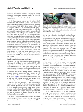
electrical power (LED: 1.5W, Ximea camera: 3.0W, and Figure 11. Reusable BARD coring needle handpiece and low-cost
a computer, which can be a battery-powered laptop), disposable needle are displayed to the photographer during a training
showing promising proof-of-concept work for low-cost course in Rwanda on breast biopsy procedure for palpable breast masses.
The photograph is provided by Dr. Jane Brock, formerly at Brigham and
and accessible solutions for rural and low-resource clinical Women’s Hospital, Harvard University.
settings. With a total cost of goods of less than USD 8,000
(excluding labor), including a camera costing USD 4,000, not yet been refined for clinical-grade imaging. Further
the system offers an affordable option compared to existing optimization is necessary to enhance contrast, reduce
digital pathology solutions. While the CoreView ION has imaging artifacts, and improve overall diagnostic quality.
not been fully automated, this was found unnecessary for Furthermore, the study has not yet demonstrated high-
achieving rapid imaging and analysis, specifically for the quality imaging of malignant BC human tissues. The
stain protocol. The simplicity and speed of the system initial results provide a foundation for future work, but
suggest that automation could easily be implemented in additional validation using a diverse range of cancerous
future iterations, but even in its current form, the workflow tissues is required to assess the system’s true diagnostic
remains efficient and practical. If further automation and potential. These limitations highlight areas for future
higher-powered LEDs were implemented, the 5-min improvement, including optimization of the staining and
process could be even faster while minimizing errors. imaging workflow, cost reduction strategies, and expanded
With increased speed provided by system automation, a validation studies to ensure clinical applicability with and
20× objective could be implemented at an incrementally without artificial intelligence (AI) enhanced diagnosis
higher cost. from the resulting CoreView ION images.
3.5. System limitations and challenges 3.6. Future improvements and optimization
While the CoreView prototype offers a promising proof- As the CoreView ION is an initial proof-of-concept
of-concept for rapid and low-cost imaging of CNBs in BC prototype, there are potential directions for further
diagnostics, several limitations must be considered before refinement of the imaging strategy and design. At present,
clinical implementation. One major limitation is the the prototype depends on a computer system for MUSE
expectation that a single core is sufficient for a diagnosis. If imaging, necessitating access to electrical power and
a second core is needed, then the needle would necessitate a computer connection. However, recent studies have
a thorough cleaning and rinsing protocol, introducing demonstrated the feasibility of utilizing MUSE imaging
potential workflow inefficiencies and requiring further through smartphones. The Pocket MUSE system, which
24
validation for sterility. An example of global use of a reusable employs an optical module attached to the rear lens of a
CNB device (Figure 11), which could be incorporated into smartphone, facilitates high-quality fluorescence imaging
a CoreView ION imaging workflow, with multiple clean at a significantly reduced cost. Incorporating Pocket MUSE
needles being used with one reusable biopsy gun.
technology and concepts into the CoreView ION could
Another issue with the system is the dependence on eliminate the requirement for a computer connection,
quartz coverslips, which are significantly more expensive enhancing its usability in rural settings. Furthermore, the
than standard glass slides. This cost factor may present existing low-powered UV LEDs in the current fixture could
a barrier to widespread adoption, particularly in low- potentially be replaced with a battery-powered module,
resource, rural settings where affordability is a primary allowing the system to operate solely on battery power.
concern. Between each sample, the quartz glass required The current prototype relies on a clean transparent
cleaning or replacing if broken, leading to workflow coverslip, which introduces workflow inefficiencies and
inefficiency between samples.
cleaning challenges. Recent advancements in imaging
In addition, the imaging workflow and staining process technologies, such as fluorescence-imitating brightfield
remain unoptimized. The current staining and imaging imaging (FIBI), have demonstrated the capability to
parameters were developed as proof-of-concept and have capture tissue images without coverslip compression. 26,27
Volume 4 Issue 3 (2025) 115 doi: 10.36922/GTM025170039