
Page 43 - IJB-2-2
P. 43
Behnam Taidi, Guillaume Lebernede, Lothar Koch, et al.
shake-flask culture in a photo-incubator (25°C) with
an atmosphere enriched in CO 2 (up to 2.0% by vol-
ume). The light at the surface of the shaken cultures
−2 −1
was 20 µmol photons m s . The rotational speed of
the orbital shaking platform was 100 rpm with a rotatio-
nal diameter of 50 cm. S. bayanus was grown in YPD
medium under the same conditions. The flasks were
only filled to 1/5 of their total volume and stoppered
with foam bungs (11901935 - X100; Fisher Scientific).
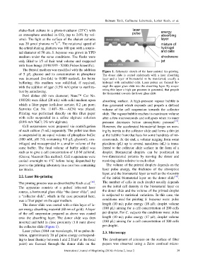
The Bristol medium was modified with the addition Figure 1. Schematic sketch of the laser-assisted bio-printing.
of 5 g/L glucose and its concentration in phosphate The donor slide is coated underneath with a laser absorbing
was increased five-fold to 0.009 moles/L for better layer and a layer of biomaterial to be transferred, usually a
buffering; this medium was solidified, if required, hydrogel with embedded cells. Laser pulses are focused thr-
with the addition of agar (1.5% w/v) prior to steriliza- ough the upper glass slide into the absorbing layer. By evapo-
tion by autoclaving. rating this layer a high gas pressure is generated, that propels
the biomaterial towards the lower glass slide.
Petri dishes (60 mm diameter; Nunc™ Cat No.
150326) were filled (20 mL) with solid medium upon absorbing surface. A high-pressure vapour bubble is
which a filter paper (cellulose acetate; 0.2 µm pore; thus generated which expands and propels a defined
Sartorius Cat. No. 11407--50----ACN) was placed. volume of the cell suspension towards the collector
Printing was performed directly on this filter paper slide. The vapour bubble reaches its maximum volume
with cells suspended in a saline alginate solution after a few microseconds and collapses when its inner
(0.9% w/v NaCl; 2% w/v alginate). pressure decreases below atmospheric pressure [15] .
Cell suspensions were prepared via centrifugation However, the accelerated biomaterial keeps on mov-
of each culture (5 mL) separately. The pellet was then ing by inertia to the collector slide and forms a thin jet
re-suspended in an equal volume of phosphate buffer at the bubble front that lasts for some hundreds of mi-
(100 mM, pH 7.0; containing 9.0 g/L glycerol); cen- croseconds. At the end, a volume ranging from some
trifuged and re-suspended in a smaller volume of the picoliters (pL) up to several nanoliters (nL) is trans-
same buffer. The final volume of buffer added was ferred to the collector slide surface in the form of a
such as to give a cell concentration of 1.0 M cells/mL droplet. Biomaterial droplets can be positioned in
(Guava; Viacount flex method). Cell suspensions were two-dimensional patterns by moving the donor and
cooled overnight to 4°C before being dispatched by receiving slides relative to each other.
post to the printing laboratory in a cool box containing The volume of the printed droplets depends on the
ice blocks. laser pulse energy, the thickness of the absorption
layer, and the biomaterial layer as well as the viscosity
2.2. Laser Bio-printing of the initial biomaterial layer on the donor slide [16] .
The printing process was as described by Koch et al. [14] . The number of cells in each droplet usually depends
The apparatus consists of a pulsed infra-red laser on the initial cell density in the biomaterial layer on
source, a horizontal glass slide “the donor slide”, and the donor slide and the volume of the printed droplet
a “collector slide”, which in the case presented here, is subjected to statistical variations. In this case, the
was a filter paper on the agar medium. conditions used for printing S. bayanus were: pulse
The donor slide was coated with a thin layer of la- length (10 ns); pulse energy (18 µJ); droplet volume
ser energy absorbing material (60 nm of gold). A layer (180 pL) aiming for a cell concentration of 200 cells
of the cell suspension prepared as above was coated per droplet. For C. vulgaris the conditions were: pulse
onto the absorbing layer. The donor slide was then length (10 ns); pulse energy (17 µJ); droplet volume
inverted and held in close proximity (1.0 mm) above (180 pL) aiming for a cell concentration of 200 cells
the collector slide (Figure 1). per droplet.
Laser pulses (1064 nm wavelength, 10 ns pulse du- 2.3. Microscopy
ration, approximately 20 µJ pulse energy correspond-
ing to laser fluency between 1 and 2 J/cm² at the focal The development of colonies on the surface of filter
point) are focused through the donor slide on the papers was observed using a Zeiss confocal micro-
International Journal of Bioprinting (2016)–Volume 2, Issue 2 39