
Page 207 - IJB-10-2
P. 207
International Journal of Bioprinting Optimizing inkjet bioprinting
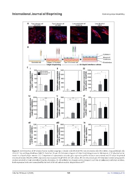
Figure 8. (A) Fabrication of 3D alveolar barrier models using type I alveolar cells (NCI-H1703), type II alveolar cells (NCI-H441), lung endothelial cells
(HULEC-5a), and lung fibroblasts (MRC5). Scale bar: 50 µm. (B) Schematic diagram of inkjet bioprinting process for fabrication of 3D alveolar barrier
model in a layer-by-layer manner. (C) Comparison of representative alveolar gene expression profiles between conventional and 3D inkjet-bioprinted
structured models. Relative mRNA expression was measured via qPCR for 2D cell culture, 3D non-structured, and 3D-structured models using specific
markers involved in (i–iii) intercellular junction formation, (iv–vi) epithelial ion channels and ion transport, and (vii–ix) pulmonary surfactant secretion.
108
Each expression levels were normalized by the level of 2D cell culture models. Adapted from ref.
Volume 10 Issue 2 (2024) 199 doi: 10.36922/ijb.2135