
Page 516 - IJB-10-2
P. 516
International Journal of Bioprinting
http://doi.org/10.18063/ijb.XXXXXXXXXXXXX
the hydrophilicity assay showed statistically significant differences (* p < 0.05) between controls and
NaOH-treated groups at each measuring point (day 0, day 1, day 2, and day 9), with a constant shift of
~9° to 15° observed between conditions. Similarly, statistically significant differences in contact angle
within time were separately found in both conditions. Additionally, the contact angle in the controls was
International Journal of Bioprinting Oozing 3D-printed scaffolds for tissue engineering
statistically significant ( p < 0.05) between day 0 and day 1, and between day 0 and day 2. However, the
FIG.6 SEM
NaOH-treated group showed differences between day 0 and day 2, and between day 0 and day 9 (Figure
8).
A B
Gy
0h
Gof
Oc
Or
Os
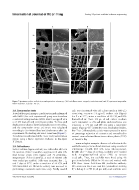
Figure 7. Specimens surface analysis by scanning electron microscopy. (A) Control specimens’ images (prior to treatment) and (B) specimens images after
Figure 7. Specimens surface analysis by scanning electron microscopy. (A) Control specimens’ images
NaOH treatment. Scale bar: 100 µm.
(prior to treatment) and (B) specimens images after NaOH treatment. Scale bar: 100 µm.
2.8. Compression tests cells were incubated with cell culture medium (600 µL)
A total of five specimens per condition (control and treated containing resazurin (10 µg/mL) sodium salt (Sigma)
with NaOH) for each experimental group were tested on for 2 h at 37°C, under a condition of 5% CO and 95%
2
a universal testing machine (Z005, Zwick) equipped with humidified air. Then, 100 µL of cell culture medium
a 2.5 kN load cell and compression plates. The load and were transferred to a 96-well plate, and absorbance was
displacement values of the tested specimens were recorded, measured at 570 nm and 600 nm using a microplate
and the compression stress and strain were calculated reader (Synergy HT Multi-detection Microplate Reader,
according to the obtained load and displacement after the Bio-Tek). Cell metabolic activity was expressed in terms
experiments. The loading rate was at 5 mm/min (Figure 9). of percentage reduction of resazurin and normalized to
E modulus was calculated at the linear elastic deformation control values obtained from tissue culture plastic (TCP)
region using a linear regression included in TestXpert of the same day.
II (Zwick).
Immunological assays to observe cell adhesion to the
2.9. Cell cultures scaffolds were performed and observed using a confocal
SaOs-2 cell line (Sigma-Aldrich) was cultured in McCoy’s microscope (CLSM, TCS SP8, Leica Microsystems).
5A medium (Fisher Scientific), supplemented with 10% Briefly, after 7 days of seeding, scaffolds were washed
fetal bovine serum (FBS, Sigma) and 1% penicillin– with phosphate-buffered saline (PBS) to remove
streptomycin (Fisher Scientific). A total of 300,000 cells dead cells. Then, the scaffolds were fixed using 4%
were seeded per scaffold. Cells were incubated for 1, 3, paraformaldehyde (PFA) for 30 min and washed with
and 7 days at 37°C, under a condition of 5% CO and PBS three times. Acti-Stain 488 phalloidin antibody
2
95% humidified air. Metabolic activity was assessed by (Cytoskeleton, Inc.) was used for cytoskeleton staining
performing a resazurin reduction assay. Briefly, seeded by diluting it in PBS (7 µL per mL), and scaffolds were
Volume 10 Issue 2 (2024) 508 doi: 10.36922/ijb.2337